Figures & data
FIGURE 1. Red-free fundus photo shows clear delineation of multiple subretinal fluid pockets and retinal folds in the posterior pole in both eyes.
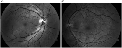
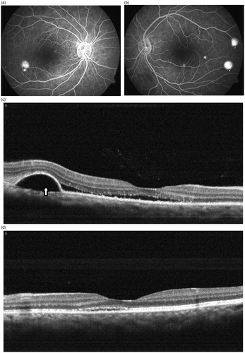
FIGURE 2. (a) Typical smokestack leak (arrow) with optic disc staining and multiple pinpoint leaks were seen at the posterior pole in the right eye. (b) Two small PED leaks (arrowheads) with disc staining and multiple pinpoint leaks are seen at the posterior pole in the left eye. (c) OCT scan in the right eye shows serous detachment of macula (measuring 958 μm), folds of RPE, and a small pigment epithelial detachment (arrow) corresponding to the smokestack leak seen on FA. (d) OCT scan in the left eye shows serous detachment of macula (measuring 208 μm), a small PED (arrowhead), and folds of RPE.
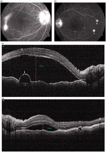
FIGURE 3. (a, b) Substantial angiographic resolution of smokestack leak (arrow) in right eye with persistence of PED leaks (arrowheads) in both eyes. (c, d) OCT scan of the right eye shows partial resolution of serous detachment of macula with persistence of PED (arrow) seen temporally and substantial resolution of serous detachment of macula seen in the left eye.