Figures & data
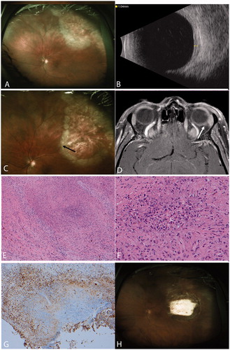
FIGURE 1. (A) Fundus photography demonstrating superotemporal area of chorioretinitis. There is a leading edge of inflammatory infiltrate with atrophy and scarring anterior to it. (B) B-scan ultrasound shows thickening of the retina and choroid. (C) Two months after her initial presentation, the area of retinitis has expanded. Note that the bifurcation of the vessels indicated by the arrow was not engulfed by the lesion in . (D) T1 post contrast fat-saturated MRI images demonstrate increased enhancement of the posterior sclera in the left eye (arrow). (E) Hematoxylin and eosin, 100× photomicrograph demonstrating granulomatous scleral inflammation. The lesion has a focal area of necrosis containing neutrophils surrounded by giant cells and epithelioid cells. (F) Hematoxylin and eosin, 200× photomicrograph shows focal area of necrosis with neutrophils. (G) Chromagranin diaminobenzine reaction, 100× photomicrograph stain for CD68 demonstrates positivity for histiocytes in the region surrounding the focal necrosis. (H) Postoperative fundus photography showing the healed biopsy site and no further progression of the chorioretinitis.