Figures & data
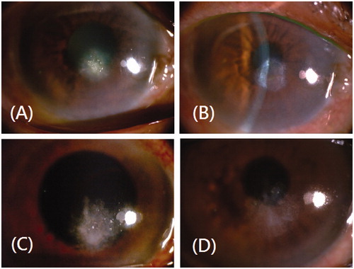
Figure 1. Case 1 presentation. (A) Slit-lamp microscopic images showing a dense, whitish, elevated inferior paracentral corneal ulcer, associated with corneal edema and localized necrosis at its periphery of the right eye cornea. (B) Fourteen days after therapy, slit-lamp microscopic images showing a quiet stromal scar with resolved corneal lesion. Case 2 presentation. (C) Slit-lamp microscopic images showing a paracentral corneal ulcer (2 × 2 mm), associated with “fluffy”-appearing infiltrates and feathered borders. (D) Fourteen days after therapy, slit-lamp microscopic images showing a faint stromal scar with corneal ulcer subsided.