Figures & data
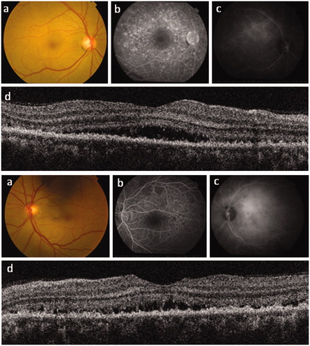
Figure 1. Right and left eye posterior segment imaging for case 1. (a) Color photography; (b) fundus fluorescein angiography (FFA); (c) autofluorescence imaging; and (d) spectral domain optical coherence tomography (OCT) imaging. Note the patchy retinal pigment epithelium loss on OCT with corresponding hyperfluorescence on FFA and hypoautofluorescence. The right OCT shows loss of the ellipsoid zone, which explains the marked reduction in visual acuity. OCT also reveals shallow serous macular detachments, particularly of the right eye.
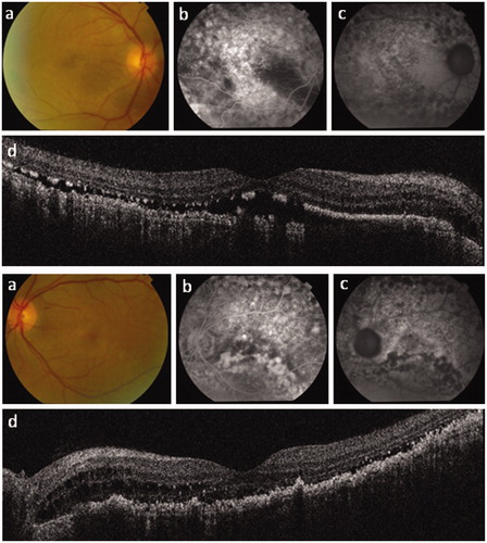
Figure 2. Right and left eye posterior segment imaging for case 2. (a) Color photography; (b) fundus fluorescein angiography; (c) indocyanine green fundal angiography; and (d) spectral domain optical coherence tomography (OCT) imaging. OCT reveals diffuse thickening of the retinal pigment epithelium (RPE) with shallow serous macular detachments bilaterally. In contrast to case 1, this patient shows no full-thickness RPE defect, with correspondingly less hyperfluorescence seen on fluorescein angiography and relatively well-preserved visual acuity. The inner retinal layers, including the papillo-macular bundle, are well preserved bilaterally.