Figures & data
Figure 1. Both fundi showing classic end stage birdshot picture; profound optic atrophy surrounded by large patches of atrophic retina, multiple deep oval lesions extending to the midperiphery and involving the macula. There is profound arteriolar attenuation and mild inactive-appearing venous sheathing. There are also multiple punched out lesions.
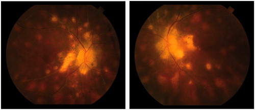
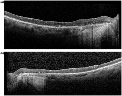
Figure 2. (a) Macular OCT OD. Loss of architecture of the inner retinal layers and diffuse atrophy of the outer layers. Residual photoreceptor layer can be seen in the foveal region. Nasal to the fovea, the RPE is atrophic (corresponding to atrophy seen in the colored fundus photograph). The choroid is visualized and appears atrophic. (b) Macular OCT OS. Shows a similar picture as OD with an irregular subfoveal photoreceptor layer. Nasal to the fovea, the RPE is atrophic.