Figures & data
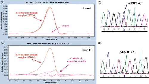
Figure 1. Mutation screening by HRM analysis and direct sequencing of PCR-amplified products. Results are shown for ITGB3 exons 5 and 11 for family members of pat 1. In panels A and B, we illustrate normalized and temperature-shifted melting curves of mutated and control PCR amplicons (Roche Light cycler 480 ResoLight Dye; Roche Diagnostics, Meylan, France) (c.685C > T, Leu196Pro; and c.1871G > A, Cys598Tyr). Control patterns (pink) mutated patterns (red) are clearly distinguished. Whereas the ITGB3 exon 5 substitution was present in all of the family members (see superimposed lines), the propositus was the only family member to possess the exon 11 mutation. In panels C and D are shown the sequencing profiles of respective heterozygous mutated PCR products. Methodological details will be supplied on request.