Figures & data
Figure 1. Illustration of tumour blood vessel fenestration due to the tumour growth and inflammatory condition in this tissue. The liposomes circulate in the blood and extravasate into the extracellular space through pores between the endothelial cells lining the blood vessel. Here the liposomes can encounter secreted enzymes such as sPLA2 or MMPs that then hydrolyze membrane moieties and induce drug release.
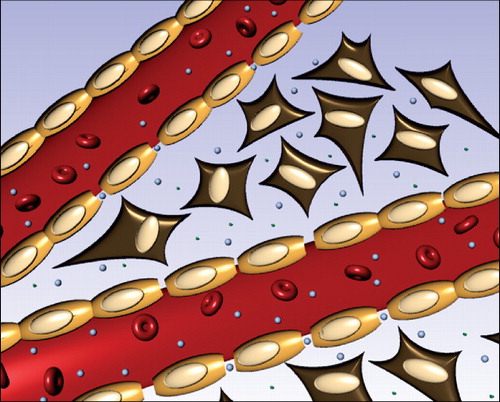
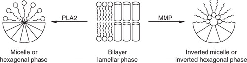
Figure 2. Illustration of how enzymes can induce a morphological change in the liposome membranes giving drug release, or induce transfection with siRNA or DNA plasmid. As an example PLA2 induces a transition to either micelle or a hexagonal phase due to the formation of lysolipids, whereas MMP will reduce headgroup size and induce a transition to an inverted micelle or the inverted hexagonal phase.