Figures & data
Figure 1. TEM images showing vesicles in various stages of membrane fusion in fossil plant cells. The membrane fusion stages are labeled by numbers. CW, cell wall. Bar = 100 nm. (a) About nine vesicles in various stages of membrane fusion in the same cell. Vesicles in S1 are free from the CM, those in S4 are already fused with the CM. Note the vesicle in the bottom right is blurry while those in the left are sharp. (b) A vesicle near the CM. Note its generally regular spherical form and a slightly distorted region (to the upper left) close to the CM. (c) A vesicle tethered to the CM. Note the cylindrical connection (arrow) between the vesicle and CM. Courtesy of Wang et al. (Citation2007) and MMB. (d) A vesicle fused with the CM. Note the two leaflets of the CM and only one leaflet of the vesicle. The vesicle membrane is connected to the inner leaflet of the CM. Courtesy of Wang et al. (Citation2007) and MMB. (e) Four vesicles in different stages of membrane fusion in the same cell. The vesicle in S2 is only weakly connected to the CM (arrow), the one in S4 is already connected to CM by a narrow neck, the one in S6 is omega-shaped and with a central plug in the fusion opening, and the one in S7 has a wider fusion opening and no visible central plug. (f) Four vesicles in different stages of membrane fusion in the same cell. Note the double layer structure of the CM and its relationship with the membranes of the vesicles. The vesicle in S1 is free from the CM, the one in S5 is already connected to the CM and forms a central plug together with the CM, the one in S6 is connected to the CM and has a relict central plug, and the one in S4 is connected to CM and has no trace of the outer leaflet of the CM. Note all vesicle membranes are connected to the inner leaflet of the CM. Rightmost, there appears to be another vesicle (arrow) with double-leaflet membrane close to its complete fusion with the CM. (g) A vesicle close to complete fusion with CM. Note its shape, relationship with the CM, and a relict plug in the fusion opening.
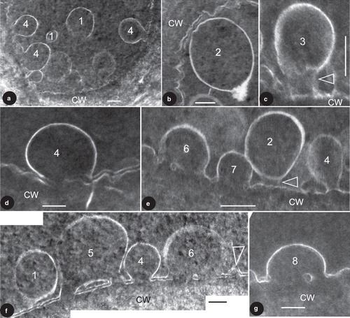
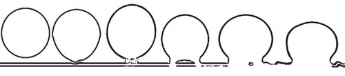
Figure 2. Diagram showing the idealized stages of membrane fusion during an exocytosis.