Figures & data
Figure 1. Network-like domains of proteins and lipids. (A) Pattern of the ATPase Pma1 in the budding yeast PM imaged by TIRFM. (B) Domains of FITC-LB21 peptide in a GUV made of PC and cholesterol. Reproduced from Kaiser et al. (Citation2011). (C) Network pattern in a GUV made from native pulmonary surfactant (NPS), with DiIC18 (red) and Bodipy-PC (green). Reproduced from Bernardino De La Serna et al. (Citation2009). (D) Patterns formed in simulations of lipid segregation by hydrophobic mismatch. Reproduced from Wallace et al. (Citation2006). Scale bars: 2 μm (A) and 10 μm (B, C). This Figure is reproduced in color in the online version of Molecular Membrane Biology.
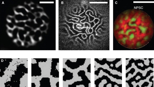
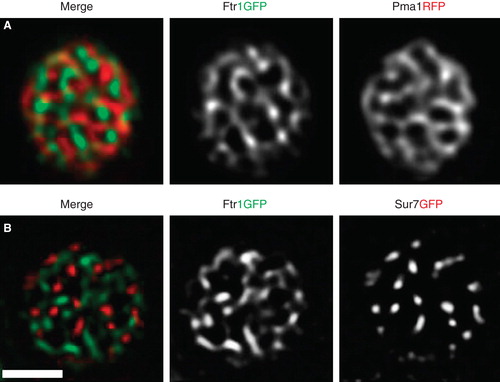
Figure 2. TIRFM of PM proteins in yeast. Images of cells transfected with Pma1RFP and Ftr1GFP (A) or Sur7RFP and Ftr1GFP (B) are shown. Both combinations show segregated membrane domains. Scale bar: 2 μm. This Figure is reproduced in color in the online version of Molecular Membrane Biology.