Figures & data
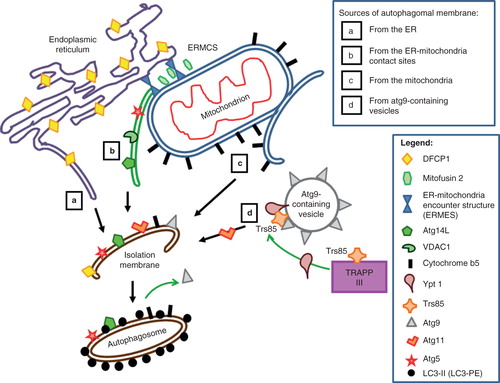
Figure 1. Possible membrane sources for forming autophagosome, or isolation membrane (IM), and the various autophagy (Atg) components and other markers associated with these. Four possible sources have been proposed to give rise to the IM. (a) Autophagosomal membrane from the endoplasmic reticulum. DFCP1, which marks the autophagosome assembly sites, is also ER-associated. (b) IM contribution from the ER-mitochondria contact site (ERMCS). Protein markers shown on this IM include the mitochondria-associated ER membrane (MAM)-enriched voltage-dependent anion channel 1 (VDAC1) and autophagy proteins Atg5 and Atg14L. The contact site is maintained by tethering molecules such as the ER-mitochondria encounter structure (ERMES) complex (Kornmann et al. Citation2009). (c) IM contribution from the mitochondria outer membrane. A mitochondrial outer membrane protein marker cytochrome b5 (cb5) is present on autophagosomes induced by starvation. (d) IM contribution from Atg9-containing vesicles. Markers such as Ypt1 and Trs85 interact on the membranes of these vesicles. Atg11, together with Ypt1, facilitates pre-autophagosome formation and recruits Atg9 to the pre-autophagosomes.