Figures & data
Figure 1. Activation of AMPK reduces NKCC1-mediated flux. Rubidium flux was measured in cells stimulated with either 1 mM AICAR or 1 mM phenformin for 1 hour. Cell types were; (A) MDCK, (B) MMDD1, (C) mIMCD3, and (D) BAEC. NKCC1 (or NKCC1/2 in the case of MMDD1 cells) mediated flux was determined as vehicle-treated flux (black bars) above the presence of 5 μM bumetanide (white bars). Fluxes were performed with n = 6 on at least three separate occasions. Counts were standardized against untreated control and then pooled. *p < 0.001.
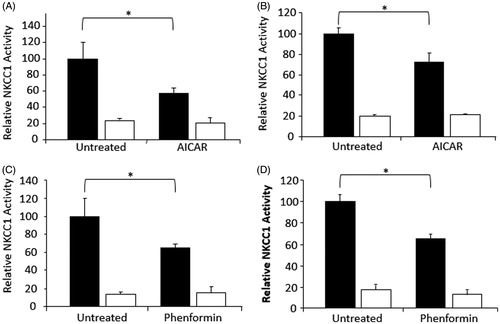
Figure 2. Activation of AMPK with A-769662 reduces NKCC1-mediated flux and phosphorylation in MDCK cells. (A) MDCK cells were stimulated with 100 μM A-769662 for 30 minutes prior to rubidium flux. NKCC1-mediated flux was determined as vehicle treated flux (black bars) above the presence of 5 μM bumetanide (white bars) (n = 24). (B) Cells stimulated with A-769662 showed marked AMPK activation (n = 3). (C) Following pre-treatment with A-769662, NKCC1-mediated flux was measured at time points for up to 60 minutes (n = 6 at each time-point). (D) Phosphorylation of NKCC1T212/217 was measured by Western blot analysis relative to β-tubulin and measured by densitometry (n = 10). *p < 0.001, **p < 0.0001 relative to vehicle-treated cells at the equivalent time-point.
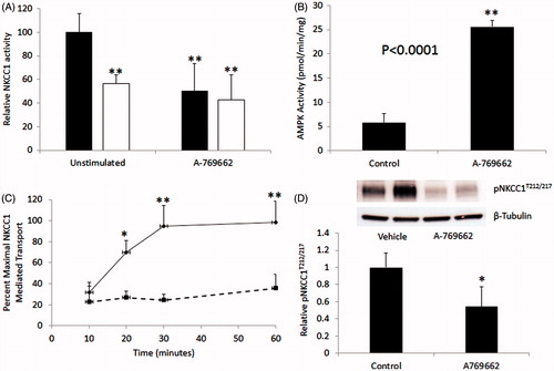
Figure 3. AMPK phosphorylates NKCC1 on S77. (A) In vitro phosphorylation of the N-terminal fragment of human NKCC1 (1–293) by purified AMPK. (B) Identification of AMPK phosphorylation site on NKCC1 by tandem mass spectrometry showing increase in the amount of 943.1(3+) AMU ion after treatment with AMPK. (C) CID spectra of the 943.1 AMU ion showing y ions and the corresponding sequence. Y5 shows loss of PO4 (18 AMU), indicating that y4 (not observed) was phosphorylated. Spectra also shows predominate neutral loss of 98 AMU (PO4) from the parent ion.
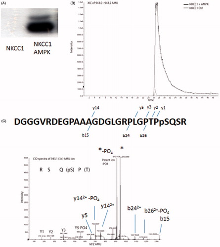
Figure 4. Mutation of NKCC1S77A results in a partial protection against AMPK-mediated reduction of NKCC1 activity. HEK293 cells stably expressing NKCC1 WT (A) or NKCC1S77A (B) were treated with 1 mM phenformin for 60 minutes (white bars) or vehicle (black bars) and then subjected to rubidium flux. NKCC1 mediated flask was considered to be the bumetanide sensitive flux. n = 12, *p < 0.001, #p < 0.05.
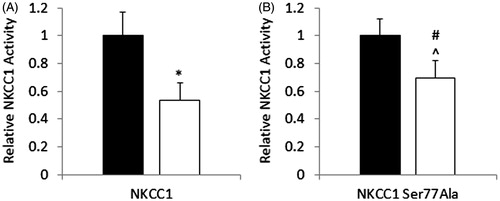
Figure 5. A-769662 mediated reduction in NKCC1 is AMPK-dependant. MDCK cells were treated with A-769662, with or without the AMPK inhibitor Compound C. (A) Western blot analysis demonstrated that A7-69662 dephosphorylation of NKCC1T212/217 was prevented by Compound C. AMPK activity was demonstrated by phosphorylation of the downstream target ACCS79 and phosphorylation of AMPKT172. β-tubulin was used as a loading control. (B) NKCC1 activity was measured as bumetanide sensitive 86Rb flux in MDCK cells (vehicle black bars, bumetanide white bars). n = 10, *p < 0.001.
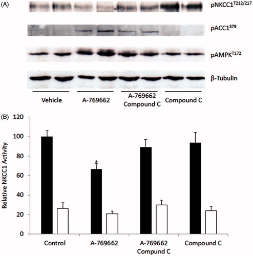
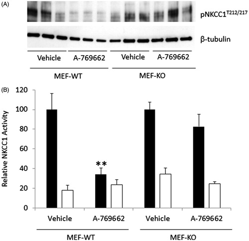
Figure 6. A-769662 mediated reduction in NKCC1-mediated flux in MEF cells is AMPK-dependent. (A) WT and AMPK null MEF cells (MEF-AMPKβ1floxβ2KO) treated with A-769662 were analyzed by Western blot to determine the phosphorylation state of NKCC1T212/217. (B) MEF cells were analyzed for NKCC1-mediated 86Rb flux (vehicle black bars, bumetanide white bars). n = 12, **p < 0.0001.