Figures & data
Table 1. Amino acid transport systems in different mammalian cells.
Figure 1. Structure of the functional chain-xCT with 12 TM domains and both the N-and C-termini are located intracellularly. Light chain is attached at TM4 by forming disulphide bond. There is a re-entrant loop in between TM2 and 3. On addition to this the binding and transporter activity is associated to TM8 (adapted from Gasol et al., Citation2004).
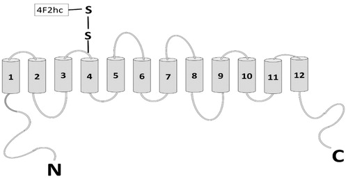
Figure 2. Nrf-2/ARE activation pathway. Under normal conditions Nrf-2 dimerizes with KEAP1 followed by its degradation due to ubiquitination by Cul3. Stimuli like free radicals and oxidizing agents can interrupt with dimerization of Nrf2 and KEAP by altering cysteine residues in KEAP1 or by phosphorylation of Nrf2 at Ser40 with the help of protein kinases. This phosphorylated Nrf2 is then translocated to the nucleus followed by its binding to the adapter proteins which increase ARE-driven transcription (adapted from Bridges et al., Citation2012b).
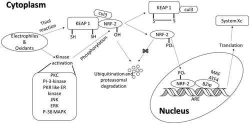
Figure 3. Production of glutathione in astrocytes and neurons via system transporter. SXc, System
, ASC, alanine-serinecysteine; MRP, multi-drug resistance proteins; GCL, glutamate cysteine ligase; GS, glutathione synthase; GSSG-GSH disulphide; TRR1, thioredoxin reductase; GGL, γ-glutamyl transferase (adapted from Lewerenz et al., Citation2013).
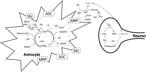
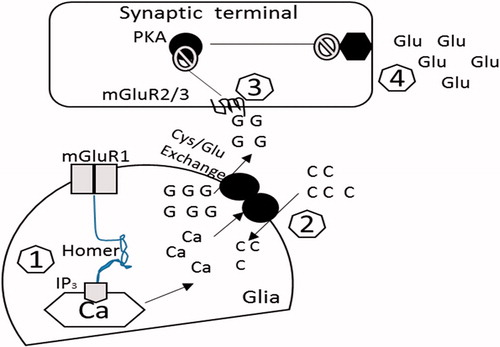
Figure 4. The possible pathways of glutamatergic transmission in the nucleus accumbens that may be responsible for drug-seeking behavior. Cocaine treatment increases the extrasynaptic release of glutamate which promotes the synaptic release of glutamate and ultimately drug-seeking behavior (adapted from Kalivas, Citation2004). G, intracellular glutamate; C, cystine; Glu, released glutamate; Ca, calcium; PKA, protein kinase A. (1) There is a reduction of homer protein in the nucleus accumbens, creating a decrease in signalling through mGluR1 receptors via inositol trisphosphate (IP3) receptor regulation of inner calcium (Ca2+) stocks. (2) The glutamate released through system activates mGlutR1. However, down-regulation of this receptor in addiction may be due to the altered activity of cystine/glutamate transporter. (3) The altered activity of system
results in the decreased release of glutamate and thus reduced activation of the presynaptic mGlu2/3 autoreceptors. (4) This decreased activation of mGlu2/3 results in a prevention of its inhibitory regulation for the glutamate release.