Figures & data
Figure 1. FCMs (ng/g) in low stereotypic (LS, n = 71–80 per day) and high stereotypic (HS, n = 69–76 per day) mink females sampled once daily at 14:00–18:00 h, given as mean ± SEM. All mink were exposed to an immobilisation challenge for 15 min at Day 2 between 09:00 and 10:00 h (indicated by the arrow). The concentration of FCM was higher in HS than in LS mink (F-test, P < 0.001), also at Day 1 (pairwise post test, P = 0.002). Significant differences from within-group baseline (Day 1) are marked with an asterisk.
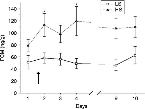
Figure 2. Response after handling without and with ACTH injection at time = 0 given as mean ± SEM concentration of (a) plasma cortisol and (b) FCM in low (LS, n = 12) and high (HS, n = 12) stereotypic mink females. Plasma cortisol increased above baseline, with no difference between LS and HS mink (F-test, P>0.53). In FCM, HS mink reacted more than LS mink during 4–20 h (F-test, P = 0.001) after handling, but not after ACTH challenge (F-test, P = 0.74). Concentrations significantly greater than baseline are marked with asterisks.
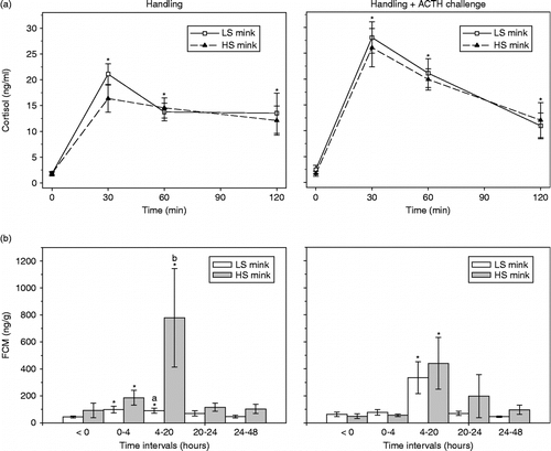
Table I. Total recovery of injected radioactivity, amount (%) excreted via the faeces, the time lag until peak concentrations of radioactivity in faeces and urine after intravenous injection of 3H-cortisol in low (LS; n = 8) and high (HS; n = 8) stereotyping female mink.
Figure 3. Time course of excretion of 3H-cortisol metabolites in (a) faeces (kBq/g) and (b) urine (kBg/ml) of low stereotypic (LS, n = 8) and high stereotypic (HS, n = 8) female mink after injection at 0 h. Data are given as hourly median with 25 and 75% quartiles for the first 16 h and per every 4th hour for the rest of the period. There was no difference between LS and HS mink for time profiles for cortisol excretion into faeces (F-test, P>0.30) or urine (F-test, P>0.50). Note the different scaling of the y-axis for faeces and urine concentration.
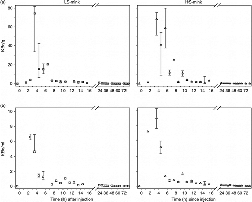
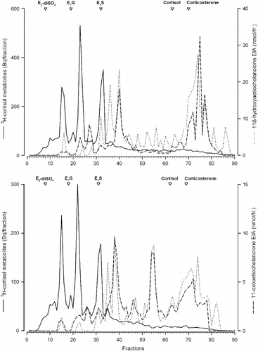
Figure 4. RP-HPLC radioimmunogram of peak radioactive faecal extracts from female mink. Samples shown are representative of a high stereotypic female (upper panel) and a low stereotypic female (lower panel) mink injected with 3H-cortisol. Radioactivity (solid line) and immunoreactivity with an 11-oxoaetiocholanolone EIA (- -) and an 11ß-hydroxoaetiocholanolone EIA (…) were determined in the different fractions. Elution times of respective standards are marked with open triangles (E2-diSO4, oestradiol disulphate; E1G, oestrone glucuronide; E1S, oestrone sulfphate, cortisol and corticosterone).