Figures & data
Figure 1. Effects of different periods of social isolation (0 min, 30 min, 2 h, 24 h and 7 days) on exploratory behavior of rats subjected to the EPM. Control rats (0 min) were kept in groups of four before being subjected to the test. Data are expressed as mean ± SEM. (A) Number of entries into the open arms of the maze; (B) number of entries into the closed arms of the maze; (C) percentage of time spent on the open arms in relation to total; (D) number of EAE; (E) number of head dips; (F) number of SAP. *p < 0.05, different from control group (0 min; one-way ANOVA followed by Newman-Keuls post-hoc test; n = 12–14 per group).
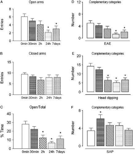
Figure 2. Plasma corticosterone concentrations (expressed as ng/ml) measured in rats subjected to different periods of social isolation (0 min, 30 min, 2 h, 24 h and 7 days) and exposed to or not to the EPM. Control rats (0 min) were kept in groups of four rats. Data are expressed as mean ± SEM. *p < 0.05, different from the respective control group (0 min); #p < 0.05, different from the group isolated for the same period and not exposed to the EPM (two-way ANOVA followed by Newman–Keuls post-hoc test (EPM exposed, n = 12–14 per group; EPM non-exposed, n = 7–8 per group).
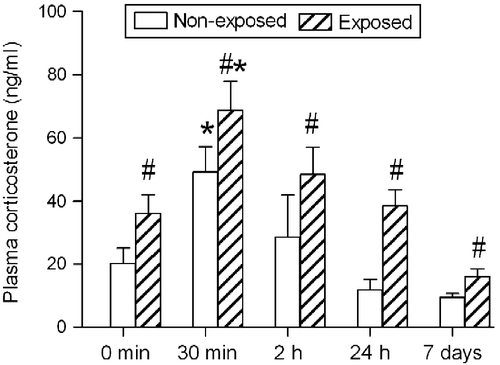
Figure 3. Spearman's correlation coefficient between plasma corticosterone concentration and frequency of SAP in rats subjected to different periods of social isolation (0 min, 30 min, 2 h, 24 h and 7 days) and exposed to the EPM. The correlation analysis included all time points of isolation, n = 66. Correlation is significant, p < 0.01.
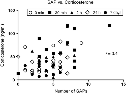
Table I. Effects of metyrapone treatment before 24 h social isolation on exploratory behavior of rats subjected to the EPM.
Figure 4. Photomicrograph of a coronal rat brain section showing representative sites and location of bilateral injection into the Cg1, based on the rat brain atlas of Paxinos and Watson (Citation2007). The number of points in the figure is less than the total number of rats used in the study because of overlapping injection sites. The positions of the representations relative to bregma are in millimeters. Scale bar = 1 mm. cc, corpus callosum.
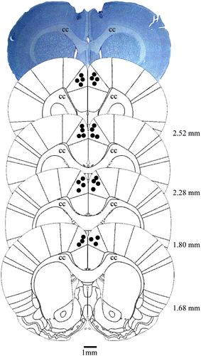
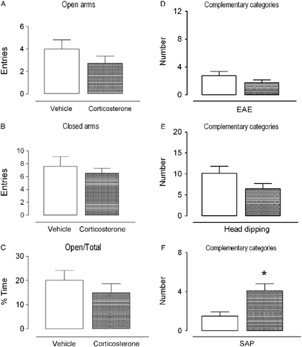
Figure 5. Effects of intra-Cg1 injection of vehicle or corticosterone (5 ng/0.5 μl) on exploratory behavior of rats subjected to the EPM. Data are expressed as mean ± SEM. (A) Number of entries into the open arms of the maze; (B) number of entries into the closed arms of the maze; (C) percentage of time spent on the open arms in relation to total; (D) number of EAE; (E) number of head dips; (F) number of SAP. *p < 0.05, different from the vehicle group (Student's t-test; n = 8–11 per group).