Figures & data
Figure 1. The effect of sex on ACTH (A) and CORT (B) concentrations at baseline (60 dB) and after 30 min of low-intensity noise stress (75 and 80 dB). All values are displayed as means ± 1 SEM. *: p < 0.05 compared to male rats at the same intensity. #: p < 0.05 compared to baseline (60 dB), regardless of sex. ^: p < 0.05 compared to 75 dB noise, regardless of sex.
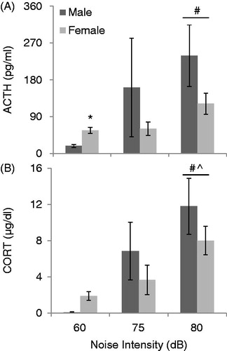
Figure 2. The effect of sex on the recovery from HPA activation in response to 30 min of 90 dB noise (black bar). All values are displayed as means ± 1 SEM. *: p < 0.05 compared to males at that time point. #: p < 0.05 compared to baseline (0 min) regardless of sex. (A) ACTH levels increased significantly in all animals after 30 min, and were back down to baseline levels by 45 minutes for both sexes. Post-hoc tests for a significant sex by time interaction revealed that males had significantly higher levels of ACTH than females at 30 min (p < 0.05). (B) CORT levels increased significantly for all animals by 30 minutes, and remained elevated until 60 minutes for both sexes. In addition, females had significantly higher baseline levels of CORT than males (p < 0.05).
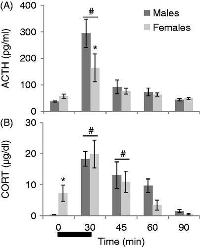
Table 1. Mean integrated densities/100 (1 SEM/100) of c-fos mRNA expression following 30 min of 90 dB noise stress.
Figure 3. The effect of sex on relative c-fos mRNA expression at baseline and following 30 min of 90 dB noise in stress-related (PL, IL, LS, BSTav, MPOA, DMH, PVN, MeA, BLA) and auditory processing (Aud Ctx, MGN, IC, VCA) brain regions. Data are expressed as mean standard (z) scores +1.0 (see “In situ hybridization” section for more information). Raw values for these data are displayed in . There was no effect of sex on either basal or stress-induced c-fos expression in any brain region examined. Abbreviations: PL: prelimbic cortex; IL: infralimbic cortex; LS: lateral septum; BSTav: bed nucleus of the stria terminalis, anteroventral subdivision; MPOA: medial preoptic area; DMH: dorsomedial nucleus of the hypothalamus; PVN: paraventricular nucleus of the hypothalamus; MeA: medial nucleus of the amygdala; BLA: basolateral nucleus of the amygdala; Aud Ctx: auditory cortex; MGN: medial geniculate nucleus of the thalamus; IC: inferior colliculus; VCA: ventral cochlear nucleus.
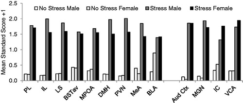
Figure 4. The effect of sex on pre-pulse suppression (GAP and PPI) and facilitation (PPF) of the acoustic startle response. All values are displayed as means ± 1 SEM. (A) No effect of sex was observed on any manipulation of the acoustic startle response (p’s > 0.05). Values represent means of the average for each animal across 5 days of testing. (B) Baseline acoustic startle amplitude (corrected for body weight (BW)) is significantly higher in female rats. Values also represent means of each animals’ average across the 5 test days. *: p < 0.05 compared to male rats. (C) No effect of sex on CORT concentration in response to 30 min of 80 dB noise was observed in the same animals.
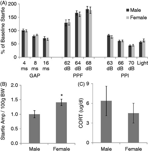
Figure 5. The effects of sex (Intact male and intact female) and ovariectomy with (OVX + E2) or without (OVX) estradiol replacement on HPA axis responses to 30 min of 105 dB noise are displayed in . Intact females were monitored for estrous cycle, but were not given stress while in a particular stage. All values are displayed as means ± 1 SEM. *: p < 0.05 compared to both intact male and OVX female rats. #: p < 0.05 compared to intact male rats. (A) ACTH concentrations before (Basal) and after noise stress. (B) CORT concentrations before (Basal) and after noise stress.
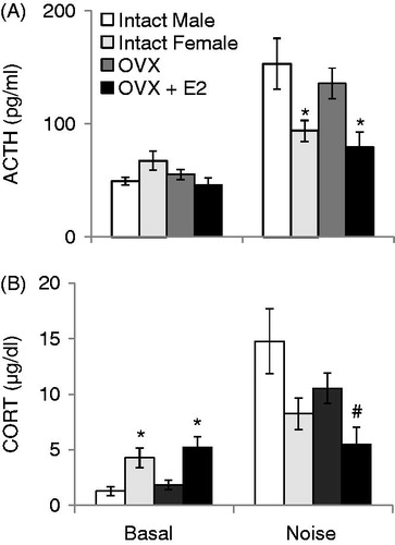
Figure 6. The effect of sex and type of stress on HPA axis responses in the same male and proestrus female rats (given restraint, then 80 dB noise, then 105 dB noise) is displayed in . Black bar represents 30 min of restraint stress. *: p < 0.05 compared to male rats at the same time point. (A) Restraint significantly increased ACTH concentrations in all animals, and female rats had significantly higher basal and stress-induced ACTH levels compared to males. (B) Restraint also increased CORT concentrations significantly. Females had significantly higher CORT levels at every time point compared to males. (C) There were no significant differences in ACTH levels due to either sex or noise intensity. (D) No effect of sex or noise intensity on CORT concentrations was observed.
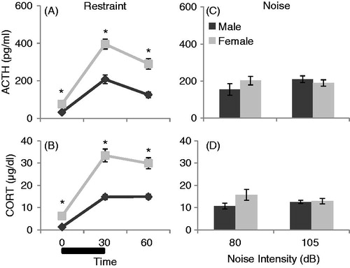
Figure 7. The effects of sex (male and females in proestrus) and social context of stress (restraint given in Groups versus in Isolation) on CORT concentrations over time (0, 30, and 60 min) are displayed in . Restraint stress for 30 min (black bar) was administered immediately after basal blood sampling performed at 0 min. (A) ACTH concentrations overall were significantly affected by stress exposure (p < 0.001) and sex (p < 0.001), but not social context (p = 0.14). Restraint stress significantly increased ACTH concentrations overall, although to a greater extent in females compared to males. (B) CORT concentrations overall were also significantly affected by stress exposure (p < 0.001) and by sex (p = 0.001), but not social context (p = 0.82). Restraint stress significantly increased CORT concentrations, and female rats had significantly higher CORT concentrations compared to males. There was also a significant time by social context interaction, where animals stressed in isolation had a significantly more prolonged response to restraint stress than animals stressed in groups, regardless of sex (p = 0.002).
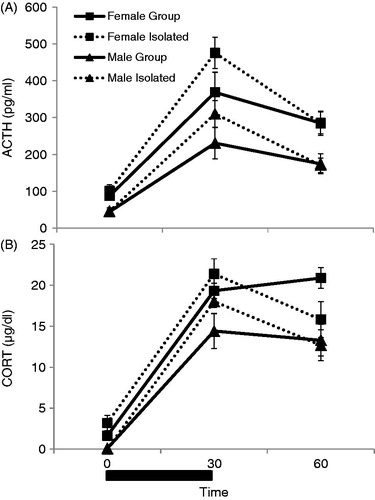
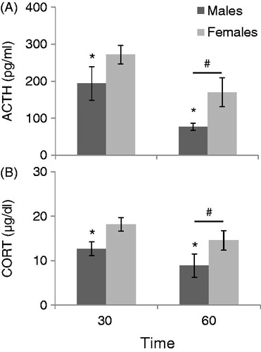
Figure 8. The effect of sex (males compared to females in proestrus) on HPA Axis responses to 30 minutes of restraint without experience of a basal blood sample is displayed in . *: p < 0.05 compared to female rats at the same time. #: p < 0.05 compared to 30 min time point. (A) Females had significantly higher ACTH concentrations than males in response to 30 minutes of restraint when one blood sample was taken either immediately after (30 min), or 30 min after being returned to the home cage (60 min). All animals overall had significantly lower ACTH levels when blood samples were taken at 60 min compared to animals that had a blood sample taken immediately following restraint (30 min). (B) Females also had significantly higher CORT concentrations than males at both time points, and all animals had significantly lower CORT levels when tested at 60 minutes than animals sampled at 30 min.