Figures & data
Table I. Description of the experimental groups of rats used in the present study.
Figure 1 Experimental design. Experimental design and groups compared for the following hypothesis: CMS induces anhedonia-like behavior and diminishes hippocampal cell proliferation and the total number of granule cells in a time-dependent manner. U-4, unchallenged 4 weeks (n = 10); U-8, unchallenged 8 weeks (n = 8); CMS-8, chronic mild stress 8 weeks (n = 8); CMS-R, chronic mild stress resilience (n = 8); and CMS-4, chronic mild stress 4 weeks (n = 10).
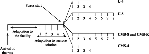
Table II. Significant differences in sucrose intake between the experimental groups.
Figure 2 Sucrose intake during the experiment. Influence of 4- and 8 weeks of chronic stress on sucrose consumption. The data were analyzed by two-way ANOVA for repeated measures, followed by Bonferroni post hoc group comparisons (). U-4, unchallenged 4 weeks (n = 10); CMS-4, chronic mild stress 4 weeks (n = 10); U-8, unchallenged 8 weeks (n = 8); CMS-8, chronic mild stress 8 weeks (n = 8), and CMS-R, chronic mild stress resilience (n = 8). Data are mean ± SEM.
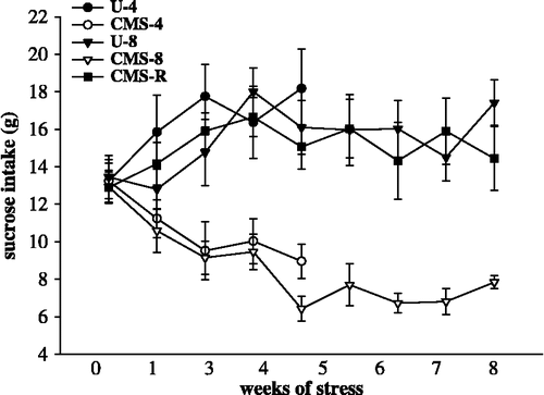
Figure 3 Cell proliferation in the hippocampal vGCL in the experimental groups. Cell proliferation in the vGCL after exposure to 4- and 8 weeks of CMS. Data were analyzed by one-way ANOVA followed by Bonferroni post hoc test. The statistical significance was set at *p < 0.05. U-4, unchallenged 4 weeks (n = 10); U-8, unchallenged 8 weeks (n = 8); CMS-4, chronic mild stress 4 weeks (n = 10); CMS-8, chronic mild stress 8 weeks (n = 8); and CMS-R, chronic mild stress resilience (n = 8). Data are mean ± SEM.
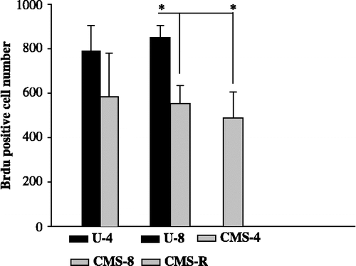
Figure 4 BrdU positive cells in the GCL of the VHF. (A) U-4, unchallenged 4 weeks, (B) CMS-4, chronic mild stress 4 weeks, (C) U-8, unchallenged 8 weeks, (D) CMS-8, chronic mild stress 8 weeks group, and (E) CMS-R, chronic mild stress resilience group. BrdU positive cells or cell clusters appear in red, the mature neurons appear in green. Scale bar: 100 μm.
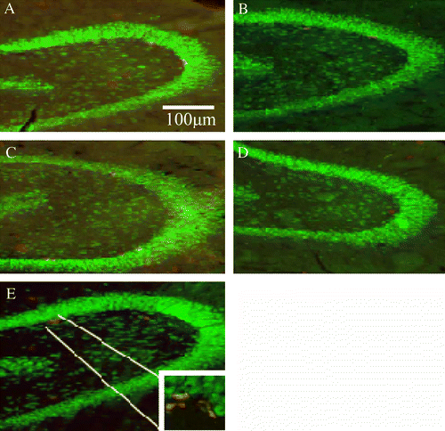
Figure 5 The total cell number in the vGCL in the experimental groups. Total cell number in the vGCL after exposure to 4- and 8 weeks of CMS. Data were analyzed by one-way ANOVA followed by Bonferroni post hoc test. The statistical significance was set at p < 0.05, *p < 0.05, **p < 0.01. U-4, unchallenged 4 weeks (n = 10); U-8, unchallenged 8 weeks (n = 8); CMS-4, chronic mild stress 4 weeks (n = 10); CMS-8, chronic mild stress 8 weeks (n = 8); and CMS-R, chronic mild stress resilience (n = 8). Data are mean ± SEM.
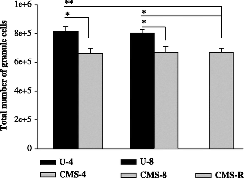
Table III. CE and CV values for estimates of the total number of granule cells in the ventral part of the hippocampal formation.
Table IV. Changes in volume of the vGCL and the ventral Ammon's horn among the experimental groups.