Figures & data
Figure 1 Behavioural responses of NS and MS rats during an open field trial. A. Mean number of faecal pellets excreted by NS (open bars, n = 10) and MS (n = 10, grey bars) rats during the 10 min exposure to open field stress. (B). Total duration spent by NS and MS rats in the inner zone (seconds, n = 5). Values are mean ± SEM. *indicates p ≤ 0.05.
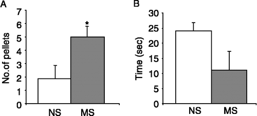
Figure 2 Behavioural responses of SD and WKY rats during an open field trial. (A). The histogram illustrates the mean number of faecal pellets excreted by SD (filled bars, n = 10) and WKY (hatched bars, n = 10) rats during the 10 min open field trial. (B). The total duration of time (seconds) spent by SD (n = 10) and WKY (n = 10) rats in the exposed inner zone of the open field arena. Values are mean ± SEM. *indicates p ≤ 0.05 and ***indicates p ≤ 0.001.
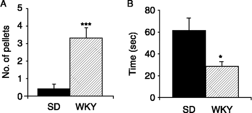
Figure 3 Goblet cell hyperplasia in MS rats. (A). The histogram shows the difference in mean colonic length (from caecum to anus) between NS (open bars, n = 5) and MS (grey bars, n = 5) rats. (B). The mean number of goblet cells per 0.5 mm muscularis mucosae in sections of the proximal and distal colon of NS and MS rats (n = 3 sections each from five rats). (C). Representative H&E stained sections from the distal colon of NS (i) and MS (ii) rats illustrate goblet cell hyperplasia in MS distal colon. Goblet cells are indicated by the arrows. Scale bar = 100 μm. Values are mean ± SEM. *indicates p ≤ 0.05, **indicates p ≤ 0.01.
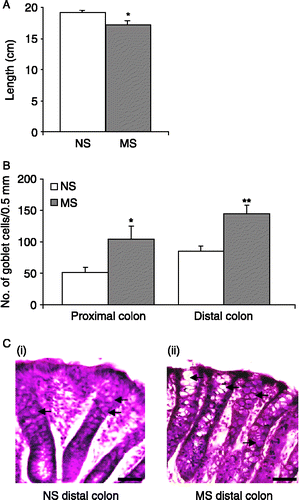
Figure 4 Thickness of the mucosal and muscular layers in MS rats. (A). The histogram illustrates the mean thickness of the mucosal layer in the proximal and distal colon of NS (n = 20 sections from four rats, open bars) and MS (n = 25 sections from five rats, grey bars) rats. (B). The mean width of the muscular layers of NS (n = 24 sections from five rats) and MS (n = 20 sections from four rats) rats in the proximal and distal colon. Values are mean ± SEM. *indicates p ≤ 0.05, **indicates p ≤ 0.01.
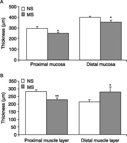
Figure 5 Goblet cell hyperplasia in WKY rats. (A). The bar chart demonstrates the small but significant increase in colonic length in WKY rats (hatched bars, n = 10) versus SD rats (filled bars, n = 10). (B). The histogram illustrates that the mean number of goblet cells in the crypts (counted for 0.5 mm of muscularis mucosae) are increased in the proximal colon of WKY rats (n = 15 sections from five rats) but unchanged in the distal colon. (C). Representative H&E stained colonic sections illustrate increased numbers of goblet cells in the proximal colon of WKY rats (ii) as compared to SD colons (i). Goblet cells are indicated by the arrows. Scale bar = 100 μm. Values are mean ± SEM. *indicates p ≤ 0.05.
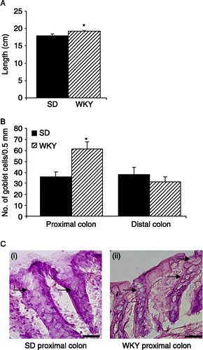
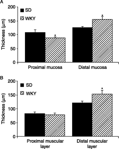
Figure 6 Thickness of the mucosal and muscular layers of WKY rats. (A). The histogram illustrates the mean thickness of the mucosal layers in the proximal and distal colon of SD (filled bars n = 25 sections from five rats) and WKYs (hatched bars n = 20 sections from four rats). (B). The mean thickness of the muscular layers in SD (n = 25 sections from five rats) and WKY (n = 20 sections from four rats) proximal and distal colons. Values are mean ± SEM. *indicates p ≤ 0.05.