1,678
Views
34
CrossRef citations to date
0
Altmetric
Review Article

Anchoring skeletal muscle development and disease: the role of ankyrin repeat domain containing proteins in muscle physiology

&
Pages 318-330 | Received 26 Feb 2010, Accepted 21 Apr 2010, Published online: 01 Jun 2010

Figures & data

Figure 1. Proposed model of evolutionary events leading to obscurin-titin binding domain (OTBD) in present-day ankyrins. In vertebrates, successive duplications led to three different modules, I, II and III. Ank1 and Ank2 have all three modules, while Ank3 has only modules I and II. Adapted from Hopitzan et al. (Citation2006). (permission has been obtained from Oxford University Press for the reproduction on this figure).
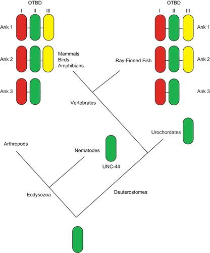
Figure 1.  Proposed model of evolutionary events leading to obscurin-titin binding domain (OTBD) in present-day ankyrins. In vertebrates, successive duplications led to three different modules, I, II and III. Ank1 and Ank2 have all three modules, while Ank3 has only modules I and II. Adapted from Hopitzan et al. (Citation2006). (permission has been obtained from Oxford University Press for the reproduction on this figure).

Table 1. Ankyrin repeat proteins expressed in skeletal muscle.

Figure 2. Caricature showing the structures in the skeletal muscle. In general, the main skeletal muscle anatomy consists of the dermomyotome, myotome and sclerotome, and is conserved throughout species. The dermomyotome is the source of the primary myotome, as well as contributing to the formation of the dermis, the endothelial and smooth muscle cells. The dermomyotome is divided into epaxial and hypaxial domains, which give rise to the epaxial muscle (deep muscle of the back) and hypaxial muscles (appendicular musculature, abdominal muscles, diaphragm, hypoglossal chords) respectively.
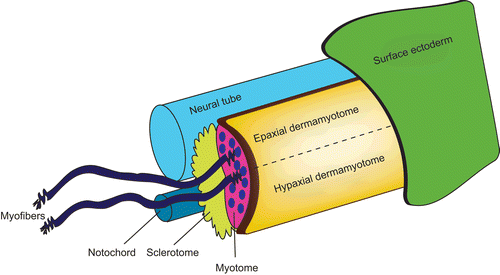
Figure 2.  Caricature showing the structures in the skeletal muscle. In general, the main skeletal muscle anatomy consists of the dermomyotome, myotome and sclerotome, and is conserved throughout species. The dermomyotome is the source of the primary myotome, as well as contributing to the formation of the dermis, the endothelial and smooth muscle cells. The dermomyotome is divided into epaxial and hypaxial domains, which give rise to the epaxial muscle (deep muscle of the back) and hypaxial muscles (appendicular musculature, abdominal muscles, diaphragm, hypoglossal chords) respectively.

Figure 3. Summary of the ankyrin repeat proteins in muscle biology, from specification and differentiation of muscle precursors (hAsb9/dAsb11, NICD, Asb15, Myo/V-1+NFΚB?, Asb2β) to the structures of the muscle fibers (MARPs).
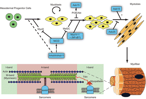
Figure 3.  Summary of the ankyrin repeat proteins in muscle biology, from specification and differentiation of muscle precursors (hAsb9/dAsb11, NICD, Asb15, Myo/V-1+NFΚB?, Asb2β) to the structures of the muscle fibers (MARPs).