Figures & data
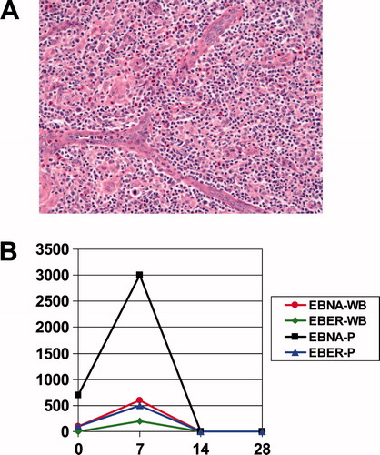
Figure 1. (A) Image from December 2001 biopsy: The nodal architecture is effaced by a polymorphic cellular infiltrate, with a spectrum of lymphoid cells (including clusters of clear cells and scattered large atypical immunoblasts), as well as eosinophils and prominent vascular elements, with frequent arborizing vessels. Hematoxylin and eosin, original magnification ×200. (B) EBV PCR on peripheral blood. Both peripheral whole blood (WB) and plasma (P) were subjected to EBV quantitative real-time PCR utilizing primer sets against EBV encoded RNA (EBER) and EBNA on days 0, 7, 14, and 28 of bexarotene (Viracor-IBT Laboratories, Lee’s Summit, MO).