Figures & data
Table 1. The composition of various formulations.
Table 2. Results of percent encapsulation efficiency, drug content, and particle size.
Figure 1. DSC thermograms of (a) pure drug, (b) physical mixture of phospholipid (DMPC) + drug, and (c) drug-loaded proliposomes (DMPC).
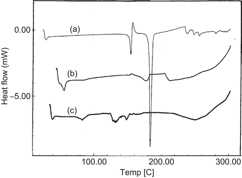
Figure 2. Series of time-lapse photomicrographs depicting the process of hydration of proliposomes (DMPC) at 400× magnification (time lapse of 1 min between each photograph).
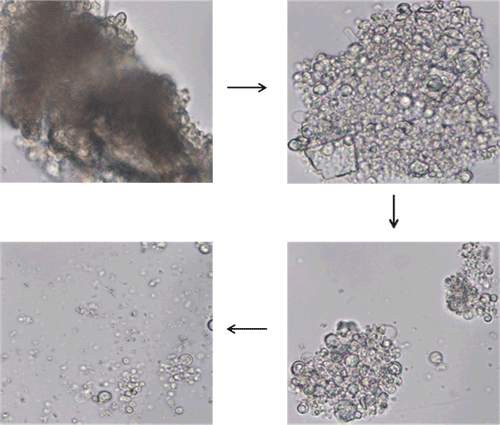
Figure 3. TEM of liposomes derived from proliposomes (DMPC).
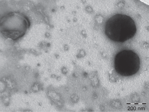
Figure 4. Photomicrograph of liposomes formed during in vitro release (4 h sample) of formulation F2 at 400× magnification.
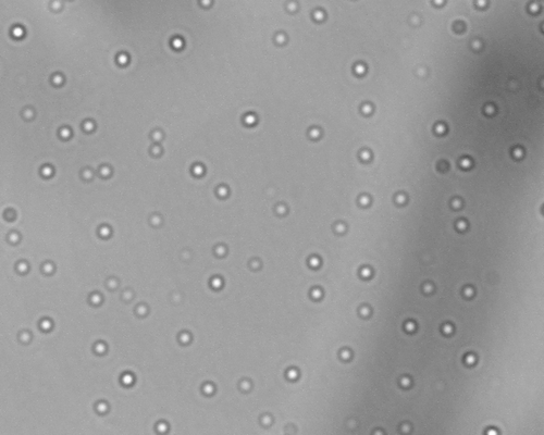
Figure 5. Evaluation of the effect of encapsulation of drug into proliposomes and incorporation of hydrophilic polymer on the in-vitro release profile of drug. Each data point represents mean ± standard deviation (n = 3).
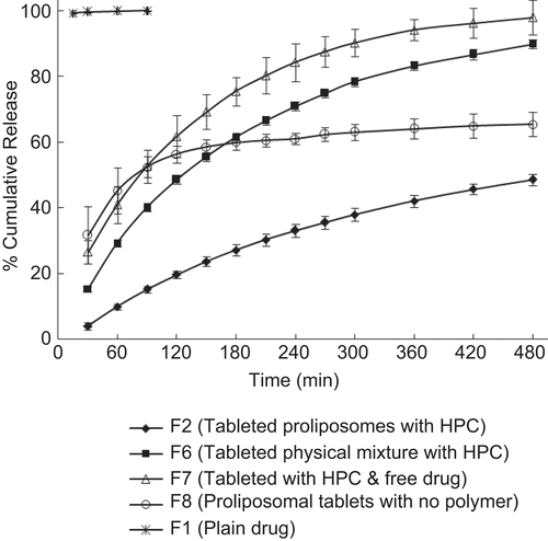
Figure 6. Illustration of possible mechanism of drug release from the proposed proliposomal multiparticulate unit dosage forms.
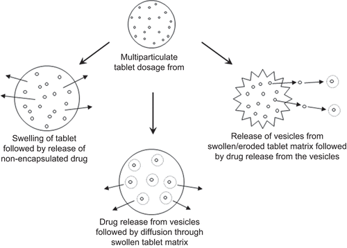
Figure 7. Effect of different phospholipid formulations on the in-vitro release profile of drug. Each data point represents mean ± standard deviation (n = 3).
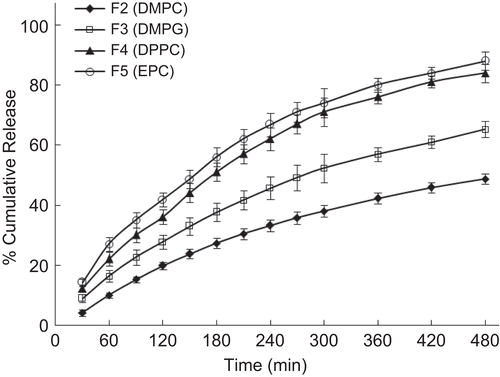
Figure 8. Effect of phospholipid concentration on the in-vitro release profile of drug. Each data point represents mean ± standard deviation (n = 3).
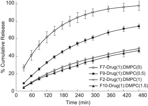
Figure 9. Effect of different polymer formulations on the in-vitro release profile of drug. Each data point represents mean ± standard deviation (n = 3).
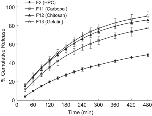
Figure 10. Effect of polymer concentration on the in-vitro release profile of drug. Each data point represents mean ± standard deviation (n = 3).
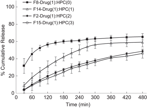
Figure 11. Effect of charge of phospholipid and polymer on the in-vitro release profile of drug. Each data point represents mean ± standard deviation (n = 3).
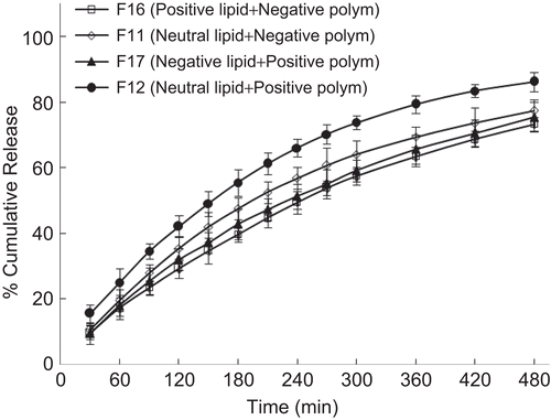
Table 3. Results of exponential coefficient (n) and correlation coefficient (r2) calculated from equation (2).