1,164
Views
36
CrossRef citations to date
0
Altmetric
Research Article

Controlled release of pentoxifylline from porous chitosan-pectin scaffolds

&
Pages 313-321 | Received 25 Nov 2009, Accepted 18 Feb 2010, Published online: 06 Apr 2010

Figures & data

Table 1. Concentrations of chitosan and pectin in the solutions used to make films and scaffolds.

Table 2. Conditions used in tests of anti-inflammatory effects of PTX release on macrophage cells in vitro.

Table 3. The decomposition temperatures, contact angles, swelling ratios, initial cell attachments (1.5 h after cell seeding), and compressional Young’s moduli of chitosan and pectin cross-linked chitosan films/scaffolds.

Figure 1. FTIR spectra of polymers made of 1% chitosan (C1), 1% chitosan and 0.5% pectin (C1P0.5), 1% chitosan and 1% pectin (C1P1), 1% chitosan and 2% pectin (C1P2), and 2% chitosan and 1% pectin (C2P1).
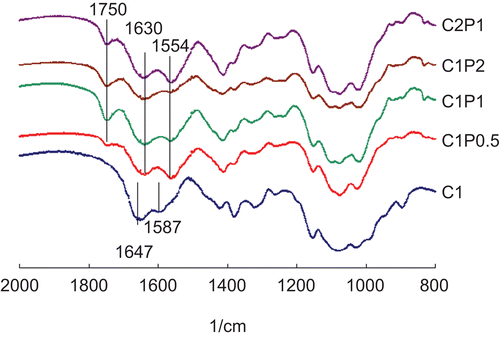
Figure 1.  FTIR spectra of polymers made of 1% chitosan (C1), 1% chitosan and 0.5% pectin (C1P0.5), 1% chitosan and 1% pectin (C1P1), 1% chitosan and 2% pectin (C1P2), and 2% chitosan and 1% pectin (C2P1).

Figure 2. Representative SEM micrographs of (a) C1 (1% chitosan), (b) C1P0.5 (1% chitosan and 0.5% pectin), (c) C1P1 (1% chitosan and 1% pectin), (d) C1P2 (1% chitosan and 2% pectin), (e) C2P1 (2% chitosan and 1% pectin) scaffolds. Scale bar = 500 μm.
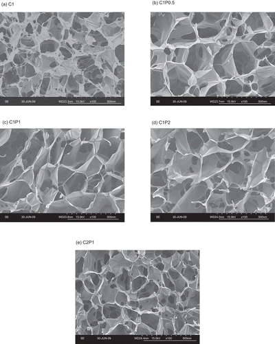
Figure 2.  Representative SEM micrographs of (a) C1 (1% chitosan), (b) C1P0.5 (1% chitosan and 0.5% pectin), (c) C1P1 (1% chitosan and 1% pectin), (d) C1P2 (1% chitosan and 2% pectin), (e) C2P1 (2% chitosan and 1% pectin) scaffolds. Scale bar = 500 μm.

Figure 3. The pentoxifylline release efficacies of 1% chitosan (C1), 1% chitosan and 0.5% pectin (C1P0.5), 1% chitosan and 1% pectin (C1P1), 1% chitosan and 2% pectin (C1P2), and 2% chitosan and 1% pectin (C2P1) scaffolds.
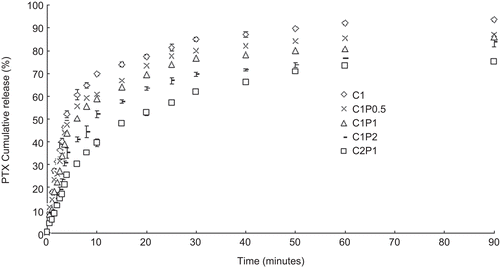
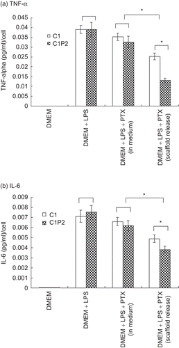
Figure 3.  The pentoxifylline release efficacies of 1% chitosan (C1), 1% chitosan and 0.5% pectin (C1P0.5), 1% chitosan and 1% pectin (C1P1), 1% chitosan and 2% pectin (C1P2), and 2% chitosan and 1% pectin (C2P1) scaffolds.
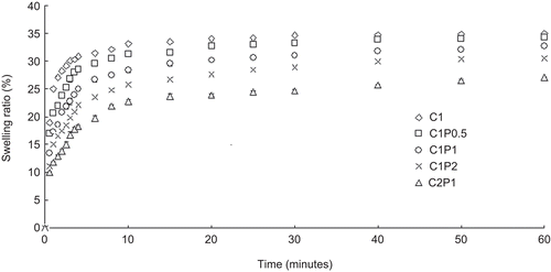
Figure 4. TNF-α (a) and IL-6 (b) release from macrophage cells cultured in the four conditions listed in . * p < 0.05.

Figure 4.  TNF-α (a) and IL-6 (b) release from macrophage cells cultured in the four conditions listed in Table 2. * p < 0.05.

Figure 5. The swelling ratios of 1% chitosan (C1), 1% chitosan and 0.5% pectin (C1P0.5), 1% chitosan and 1% pectin (C1P1), 1% chitosan and 2% pectin(C1P2), and 2% chitosan and 1% pectin (C2P1) scaffolds.

Figure 5.  The swelling ratios of 1% chitosan (C1), 1% chitosan and 0.5% pectin (C1P0.5), 1% chitosan and 1% pectin (C1P1), 1% chitosan and 2% pectin(C1P2), and 2% chitosan and 1% pectin (C2P1) scaffolds.

Reprints and Corporate Permissions

Please note: Selecting permissions does not provide access to the full text of the article, please see our help page How do I view content?

To request a reprint or corporate permissions for this article, please click on the relevant link below:

Academic Permissions

Please note: Selecting permissions does not provide access to the full text of the article, please see our help page How do I view content?

Obtain permissions instantly via Rightslink by clicking on the button below:

If you are unable to obtain permissions via Rightslink, please complete and submit this Permissions form. For more information, please visit our Permissions help page.