Figures & data
Figure 1. Plasma insulin concentration-time curve after nasal administration of chitosan hydrochloride, TMC-L and TMC-H at a concentration of 0.25% w/v (a) and at a concentration of 0.5% w/v (b) at pH 4.40 and at 0.5% w/v at a of pH7.4 (c). Each point represents the mean of six experiments.
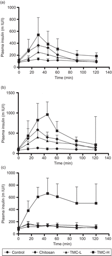
Figure 2. Plasma insulin concentration-time curve after rectal administration of chitosan hydrochloride, TMC-L and TMC-H (0.5% w/v) at pH 4.40 (a) and pH 7.40 (b). Each point represents the mean of six experiments.
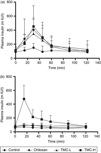
Table 1. Histological effects of chitosan hydrochloride, TMC-L, and TMC-H (0.5% w/v) on nasal and rectal epithelia.
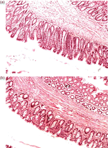
Figure 3. The effect of chitosan hydrochloride on rectal epithelia. (a) Untreated rectal epithelia. (b) Rectal epithelia after exposure (60 min) to 0.5% w/v TMC-H at pH 7.40. Note the mild congestion and edema in the submucosa (indicated by the double arrow) and the slight flattening of the epithelial cells (indicated by the arrow).