1,286
Views
21
CrossRef citations to date
0
Altmetric
Research Article

Nanoparticle-based immunopotentiation via tetanus toxoid-loaded gelatin and aminated gelatin nanoparticles

, &
Pages 320-330 | Received 02 Aug 2010, Accepted 30 Nov 2010, Published online: 25 Feb 2011

Figures & data

Table 1.   The average particle size, EE, PDI, and percentage amine group content of unlabeled gelatin nanoparticles and FITC-TT loaded GNPs and AGNPs. Values are expressed as mean ± SD (n = 3).

Figure 1.  SEM image showing spherical morphology of TT loaded AGNPs

Figure 1.  SEM image showing spherical morphology of TT loaded AGNPs

Figure 2.  Cumulative % release of FITC-TT from various formulations at various time periods up to 48 h
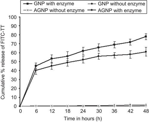
Figure 2.  Cumulative % release of FITC-TT from various formulations at various time periods up to 48 h

Figure 3.  Far-UV CD spectra of tetanus toxoid in acetone to water ratio of 1:3 at 40oC after 5h of incubation period.
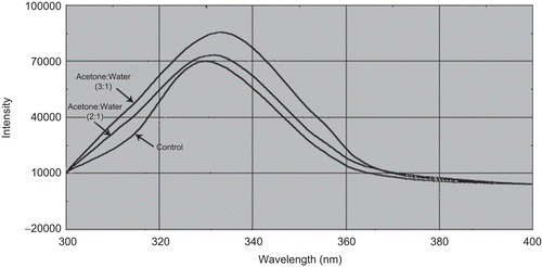
Figure 3.  Far-UV CD spectra of tetanus toxoid in acetone to water ratio of 1:3 at 40oC after 5h of incubation period.

Figure 4.  Fluorescence spectra of tetanus toxoid in acetone to water ratio of 1:3 at 40oC after 5h of incubation period.
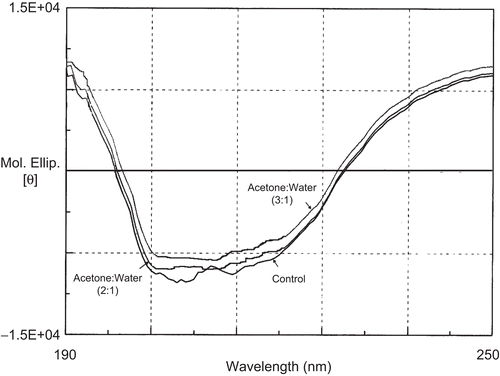
Figure 4.  Fluorescence spectra of tetanus toxoid in acetone to water ratio of 1:3 at 40oC after 5h of incubation period.

Figure 5.  Serum anti-TT profile of mice immunized subcutaneously with different formulations. Values are expressed as mean±S.D. (n = 6) 55x35mm (300 × 300 DPI)
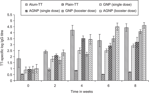
Figure 5.  Serum anti-TT profile of mice immunized subcutaneously with different formulations. Values are expressed as mean±S.D. (n = 6) 55x35mm (300 × 300 DPI)

Figure 6.  Log IgG1 and Log IgG2a titre of different formulations after s.c injection at 0 and 8 weeks. Values are expressed as mean±S.D. (n = 6) 177×105mm (72 × 72 DPI)
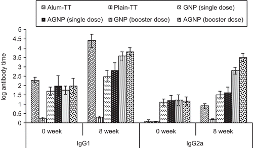
Figure 6.  Log IgG1 and Log IgG2a titre of different formulations after s.c injection at 0 and 8 weeks. Values are expressed as mean±S.D. (n = 6) 177×105mm (72 × 72 DPI)

Figure 7.  Gelatin and aminated gelatin specific IgG antibody response with selected formulation loaded with TT at 0 and 8 week following s.c administration. Values are expressed in terms of log10 IgG antibody titers 73 × 48mm (200 × 200 DPI)
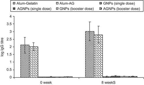
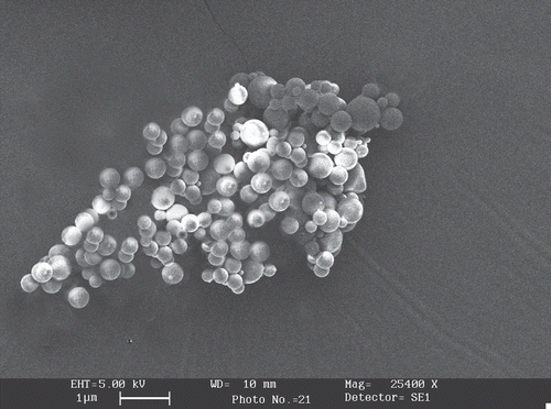
Figure 7.  Gelatin and aminated gelatin specific IgG antibody response with selected formulation loaded with TT at 0 and 8 week following s.c administration. Values are expressed in terms of log10 IgG antibody titers 73 × 48mm (200 × 200 DPI)
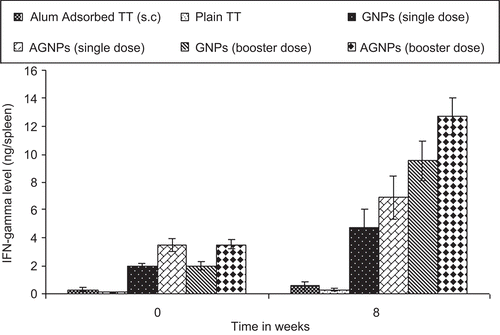
Figure 8.  Interferon-γ levels in the spleen of mice immunized with different formulations. Values are expressed as mean±S.D. (n=6) 47 × 31mm (300 × 300 DPI)

Figure 8.  Interferon-γ levels in the spleen of mice immunized with different formulations. Values are expressed as mean±S.D. (n=6) 47 × 31mm (300 × 300 DPI)

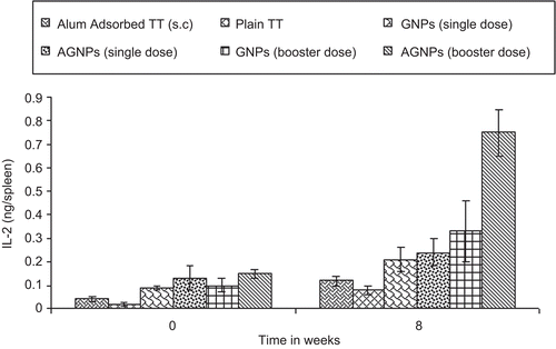
Figure 9.  IL-2 level (ng/spleen) in spleenocytes supernatant at 0 and 8 weeks after s.c injection 49×32mm (300 × 300 DPI)

Figure 9.  IL-2 level (ng/spleen) in spleenocytes supernatant at 0 and 8 weeks after s.c injection 49×32mm (300 × 300 DPI)

Reprints and Corporate Permissions

Please note: Selecting permissions does not provide access to the full text of the article, please see our help page How do I view content?

To request a reprint or corporate permissions for this article, please click on the relevant link below:

Academic Permissions

Please note: Selecting permissions does not provide access to the full text of the article, please see our help page How do I view content?

Obtain permissions instantly via Rightslink by clicking on the button below:

If you are unable to obtain permissions via Rightslink, please complete and submit this Permissions form. For more information, please visit our Permissions help page.