Figures & data
Table 1. Composition of calcium loaded BSA microspheres.
Table 2. Physicochemical characteristics of calcium loaded BSA microspheres.*
Figure 1. Scanning electron microscopy pictures of calcium loaded BSA microspheres (formulation F4) at magnification ×7000.
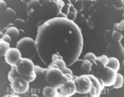
Figure 2. In vitro release profiles of calcium loaded BSA microspheres in pH 1.2 for 2 h followed by pH 6.8 for 22 h. The data represents the mean ± SD of six determinations.
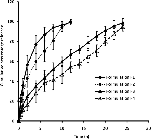
Figure 3. Serum calcium profiles obtained after oral administration of calcium loaded BSA microspheres (F4) and control solution (~1 mL, dose of 30 mg/kg of calcium) in Sprague–Dawley rats. The data represent mean ± SD of six determinations.
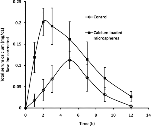
Table 3. Mean pharmacokinetic parameters of calcium in serum following oral administration of calcium loaded microspheres (F4) and control (~1 mL, dose of 30 mg/kg of calcium) for a period of 12 h in Sprague–Dawley rats (n = 6). Cmax indicates maximum concentration; Tmax, time of maximum concentration; Kel, elimination rate constant; AUC0–α, area under the serum concentration-time curve; T1/2, elimination half-life.