Figures & data
Figure 1. Viscosity-shear rate profiles of CsA emulgel.
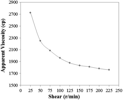
Table 1. The stability of CsA emulgel.
Figure 2. The effects of concentration and viscosity of gel on the release behavior of CsA emulgel.
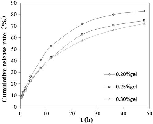
Figure 3. Concentration–time profiles of CsA in tear fluid after instillation in conscious rabbits (n = 5).
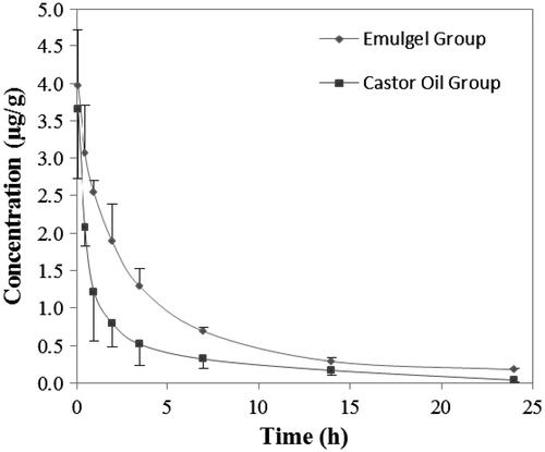
Table 2. Parameters based on statistical moment theory for CsA in tear fluid after topical administration in conscious rabbits (n = 6).
Table 3. Mean tear concentration of CsA after instillation of E-gel preparation and castor oil preparation in rabbit(mg/ml,  , n = 6).
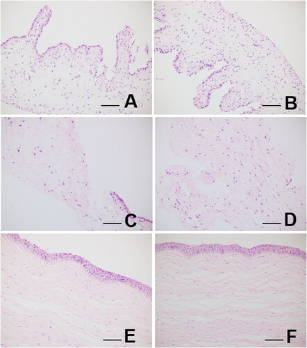
Figure 4. Light microscopy of rabbit’s iris (A, B), rabbit’s conjunctiva (C, D), and rabbit’s corneal tissue sections (E, F), from CsA emulgel-treated (OD) and control (OS) rabbit eyes. Samples and tissue sections from treated eyes showed no alterations in morphological details compared to control eyes. Eye tissues from CsA emulgel-treated animals gave similar results. Staining: (A–F) hematoxylin-eosin. Scale bar = 100 μm.