Figures & data
Table 1. Loading efficacy of different formulations.
Figure 1. IR Spectrum of curcumin (a) and curcumin-β-cyclodextrin complex (b).
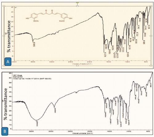
Table 2. Optimization of gelatin, formaldehyde and curcumin-β-cyclodextrin complex concentration.
Figure 2. A photomicrograph of 5% gelatin stable foam (a) and curcumin-β-cyclodextrin-loaded gelatin sponge (b) at 100×s.
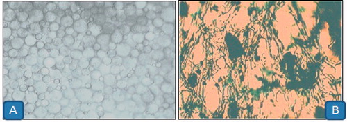
Figure 3. In vitro release of drug from sponge containing formaldehyde and without containing formaldehyde.
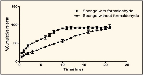
Figure 4. Histopathology of (a) plain cyclodextrin-treated group, (b) gelatin gel-treated group, (c) plain curcumin-treated group, (d) silver sulfadiazine-treated group and (e) curcumin-β-cyclodextrin sponge-treated group.
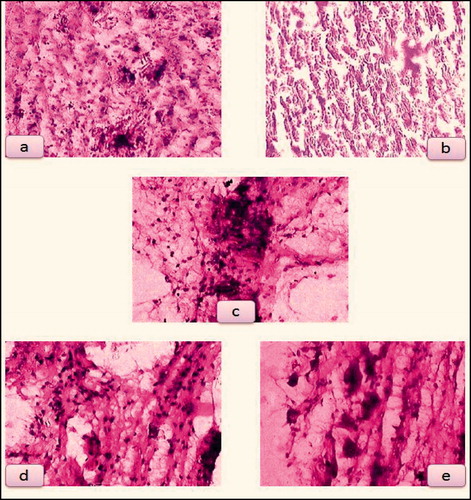
Figure 5. (a) Plain cyclodextrin-treated group, (b) gelatin gel-treated group, (c) plain curcumin-treated group, (d) silver sulfadiazine-treated group and (e) curcumin-β-cyclodextrin-loaded sponge treated group.
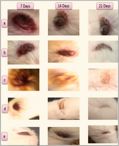
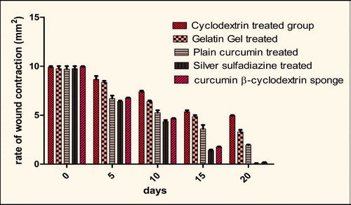
Figure 6. Changes in area of wound contraction during the progress of wound healing.