Figures & data
Table 1. Composition of the gel formulations.
Table 2. Results of pH and viscosity values of the gel formulations.
Table 3. Mucoadhesion values of the gel formulations using texture analyzer with rat colonic mucosa (n = 6).
Figure 1. Release profiles of melatonin from gel formulations. The data represent the mean ± confidence interval, n = 6.
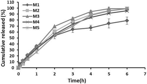
Table 4. Kinetic assessment of the release data of melatonin from gel formulations.
Table 5. The stability test results of the M5 gel formulation.
Figure 2. MDA levels in colonic mucosa. The data represent the mean ± SD, n = 6 (*p < 0.01, versus to Group 1).
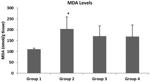
Figure 3. NOx levels in colonic mucosa. The data represent the mean ± SD, n = 6 (*p < 0.05, versus to Group 1 and Group 3, *p < 0.01, versus to Group 4).
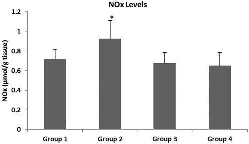
Figure 4. GSH levels in colonic mucosa. The data represent the mean ± SD, n = 6.
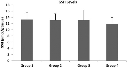
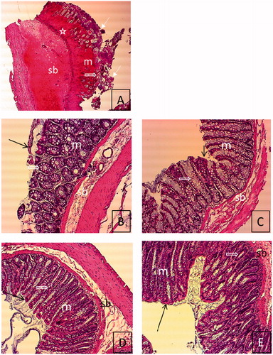
Figure 5. Histological features of rat colon sections in different groups. (A) Tissue image of group 0. (B) Tissue image of group 1, (C) Tissue image of group 2, (D) Tissue image of group 3, (E) Tissue image of group 4. Degeneration of epithelium and intestinal crypt, severe inflammatory cell infiltration, mucosal and submucosal edema in group 0 (A) (H&E. 4 × 10). Improvement in the structure of the intestinal crypt, incompleted epitelization, no edema in mucosa and submucosa in group 1 (B) (H&E. 10 × 10). Regular epithelium, improvement of the structure of the intestinal crypt, and no edema in mucosa and submucosa in Groups 2–4 (C–E) (H&E. 10 × 10). Epithelium (![]()