Figures & data
Figure 1. Distribution of non-liposomal hydrophilic dye in treated eye at three time points post-instillation. CE – corneal epithelium; CB – ciliary body; RGC – retinal ganglionic cell layer; INL/IPL – inner nuclear and inner plexiform layer; ONL/OPL – outer nuclear and outer plexiform layer; OS – outer segment.
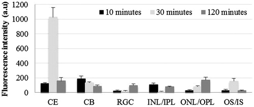
Figure 2. Distribution of hydrophilic dye used in liposomal formulation in treated eye at three time points post-instillation. CE – corneal epithelium; CB – ciliary body; RGC – retinal ganglionic cell layer; INL/IPL – inner nuclear and inner plexiform layer; ONL/OPL – outer nuclear and outer plexiform layer; OS – outer segment.
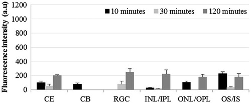
Figure 3. Distribution of liposomal hydrophilic dye in contralateral eye at three time points post-instillation. CE – corneal epithelium; CB – ciliary body; RGC – Retinal ganglionic cell layer; INL/IPL – inner nuclear and inner plexiform layer; ONL/OPL – outer nuclear and outer plexiform layer; OS – outer segment.
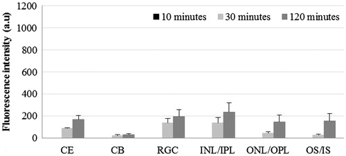
Figure 4. Confocal microscopic images of hydrophilic dye distribution in the eye at three time points (30, 60 and 120 min post-instillation) after single drop application. CE – corneal epithelium; CB – ciliary body.
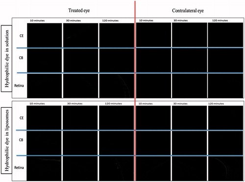
Figure 5. Distribution of non-liposomal lipophilic dye in treated eye three time points post-instillation. CE – corneal epithelium; CB – ciliary body; RGC – Retinal ganglionic cell layer; INL/IPL – inner nuclear and inner plexiform layer; ONL/OPL – outer nuclear and outer plexiform layer; OS – outer segment.
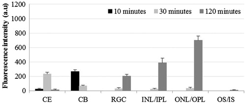
Figure 6. Distribution of liposomal lipophilic dye in treated eye at three time points post-instillation. CE – corneal epithelium; CB – ciliary body; RGC – Retinal ganglionic cell layer; INL/IPL – inner nuclear and inner plexiform layer; ONL/OPL – outer nuclear and outer plexiform layer; OS – outer segment.
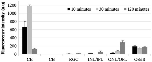
Figure 7. Distribution of liposomal lipophilic dye in contralateral eye at three time points post-instillation. CE – corneal epithelium; CB – ciliary body; RGC – Retinal ganglionic cell layer; INL/IPL – inner nuclear and inner plexiform layer; ONL/OPL – outer nuclear and outer plexiform layer; OS – outer segment.
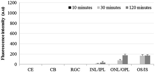
Figure 8. Distribution of non-liposomal lipophilic dye in contralateral eye at three time points post-instillation. CE – corneal epithelium; CB – ciliary body; RGC – Retinal ganglionic cell layer; INL/IPL – inner nuclear and inner plexiform layer; ONL/OPL – outer nuclear and outer plexiform layer; OS – outer segment.
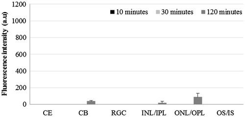
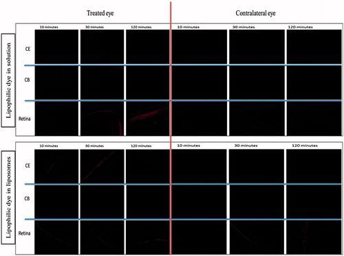
Figure 9. Confocal microscopic images of lipophilic dye distribution in the treated and contralateral eye at three time points (30, 60 and 120 min post-instillation) after single drop application. CE – corneal epithelium; CB – ciliary body.