Figures & data
Figure 1. In vitro cell variability of NP, 5-FU and 5-FU on HCT-116 colorectal cancer cell lines.
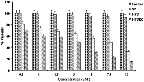
Figure 2. Nuclear morphological changes of HCT-116 colorectal cancer cell lines after 48 h by DAPI staining. 5-FUEC nanoparticles had shown morphological changes like nuclear condensation and irregular edges that can be observed in the third row of the figure (vertical).
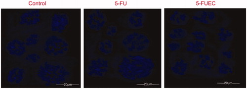
Figure 3. Apoptotic analysis of HCT-116 colorectal cancer cell lines treated with 5-FU and 5-FUEC by Annexin V-FITC/PI staining at 48 h. First row (vertical) is control that showed more than 93% viability.
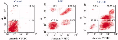
Figure 4. Fluorescent microscopic images of HCT 116 colon cancer lines with enteric coated nanoparticles, pure drug (5-FU) at 24 h and 72 h. Control was visualized by green fluorescence at all time intervals conforming its viability. 5-FU had shown minimal fluorescence of light orange and green representing minimal apoptosis. 5-FUEC had shown appreciable apoptosis attributing to the major part of the cells with red fluorescence.
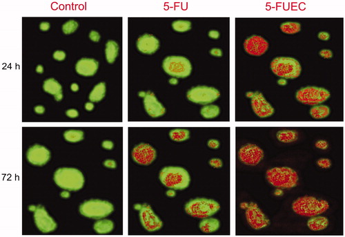
Figure 5. Mitochondria membrane potential (ΔΨm) change evaluation in HCT-116 cell lines induced by 5-FU and 5-FUEC by staining the cells with JC-1 followed by analyzing them in fluorescence microscope. (A) Control cells producing red fluorescence, (B) 5-FU (pure drug) producing partial green fluorescence representing minimal potential change and (C) 5-FUEC producing full green fluorescence representing complete mitochondrial potential change. Second and third boxes represent apoptotic activity of 5-FU and nanoparticles respectively.
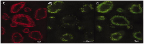
Figure 6. Visualization of release of enteric coated nanoparticulate pellets in various regions of GIT at different time intervals using γ-scintigraphy. (A) Intactness of the pellet was found unaffected in stomach (<2 h). (B) Minimal tracer release in small intestine (<2 h). (C) Release of tracer in ascending colon (after 4 h). (D) Distortion of pellet by releasing the tracer with time as it progresses in ascending colon (9 h). (E) After 12 h, the major amount of tracer release in ascending colon, transverse colon followed by pellet entry into whole colon with complete disintegration (radioactivity in distal part of colon) with increase in time till 24 h. Red color boxes indicate the γ-scintigraphic image of the pellets, where as black dotted boxes highlights the pellets shape with enhanced magnification.
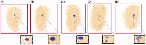
Figure 7. Tumor growth curves with various 5-FU formulation treatments (saline, NP, 5-FU, 5-FUPNP and 5-FUEC). Data are presented as mean ± SD (n = 5), p < 0.05.
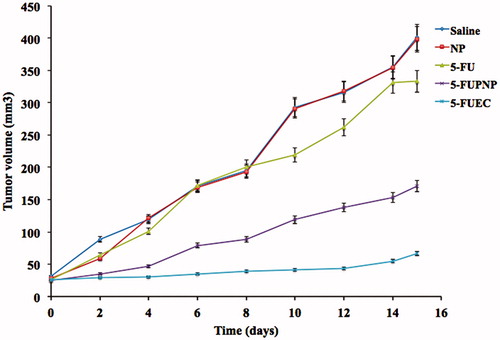
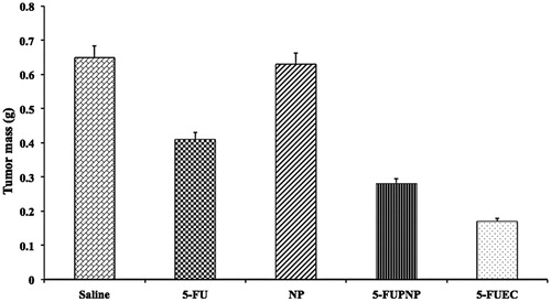
Figure 8. Measurement of tumor weights after excising the tumor xenografts by 15 days. Data are presented as mean ± SD (n = 5), p < 0.05.