Figures & data
Table 1. Primers used in this study.
Figure 1. PEAL NPs were spherical and well distributed, with no aggregation (A, left) and positively charged (A, right). EGF-PEAL NPs were also spherical (B, left) and positively charged (B, right).
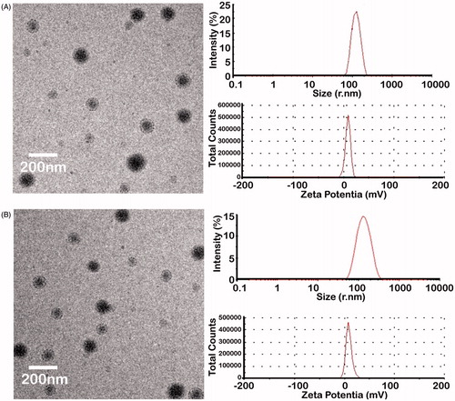
Table 2. The characterization of PEAL and EGF-PEAL.
Figure 2. Dox and siRNA release profile in vitro. (A) Dox release from free Dox, Dox-PEAL NPs and Dox-EGF-PEAL NPs. (B) Bcl-2-siRNA release from free Bcl-2-siRNA, Bcl-2-PEAL NPs and Bcl-2-EGF-PEAL NPs. Data were expressed as mean ± SD (n = 3).
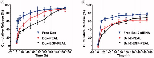
Figure 3. The cell viability of H1299 cells (A) after incubation with PEAL NPs and EGF-PEAL NPs of different concentrations for 24 h, 48 h and 72 h or (B) incubated with free Dox or Bcl-2 siRNA or EGF-PEAL NPs loaded Dox or siRNA (Dox 1 μg/ml; siRNA 50 nm).
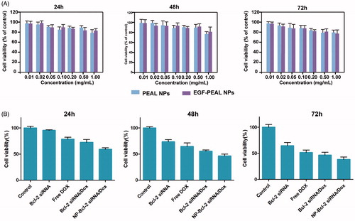
Figure 4. The uptake of Dox and FAM-Bcl-2 siRNA which were co-delivered by EGF-PEAL NPs, by H1299 at different points (1 and 6 h). At 1 h, compared to control group (free Dox and free FAM-Bcl-2 siRNA, 1 h), the drug and gene co-delivery group increased the uptake rate of Dox and siRNA. As the time of incubation increased (6 h), the enhancing effect became more obvious.
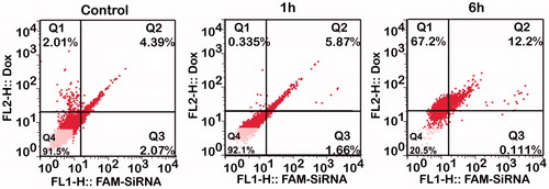
Figure 5. The results of real-time PCR, which detected the gene silencing effects of free Bcl-2-siRNA, Bcl-2-PEAL NPs and Bcl-2-EGF-PEAL NPs. The effects of Bcl-2-PEAL NPs and Bcl-2-EGF-PEAL NPs were better than free Bcl-2-siRNA (#p < 0.05, *p < 0.05). All bars represent mean ± SD (n = 3).
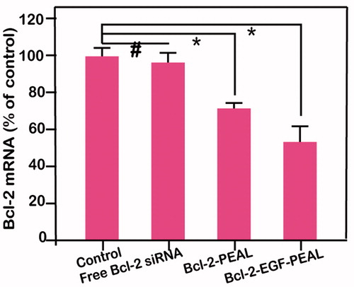
Figure 6. Apoptotic effect of H1299 cells after incubation with Dox-EGF-PEAL NPs, Bcl-2-EGF-PEAL NPs, free Dox and free Bcl-2 siRNA for 24 h.
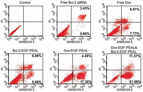
Figure 7. Distribution of Cy5-siRNA-PEAL NPs and Cy5-siRNA-EGF-PEAL NPs in vivo. (A) Images of Cy5-siRNA-PEAL NPs and Cy5-siRNA-EGF-PEAL NPs at 6, 24 and 48 h after being injected from tail veins in vivo. (B) The fluorescent images of hearts, livers, spleens, kidneys and tumors after 48 h. (C) Cryostat sections of tumor tissues. The fluorescent intensity of Cy5-siRNA-EGF-PEAL NPs in the tumor was obviously stronger than Cy5-siRNA-PEAL NPs group.
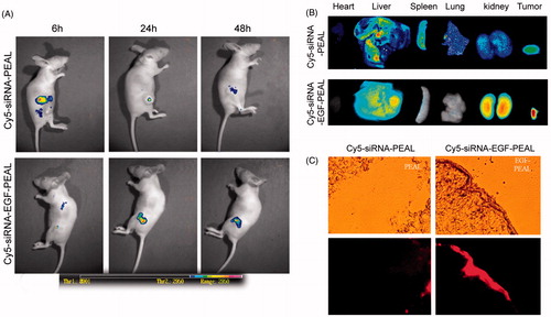
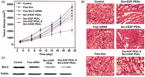
Figure 8. Therapeutic effect of EGF-PEAL co-delivery Dox and Bcl-2-siRNA on H1299 transplanted tumors. (A) The growth curve of tumors in different groups. (B) The morphological features of H1299 transplanted tumors in different groups. (C) The expression of Bcl-2 protein in H1299 transplanted tumors.