Figures & data
Figure 1. The solubility of AmB in various lipids and surfactants.
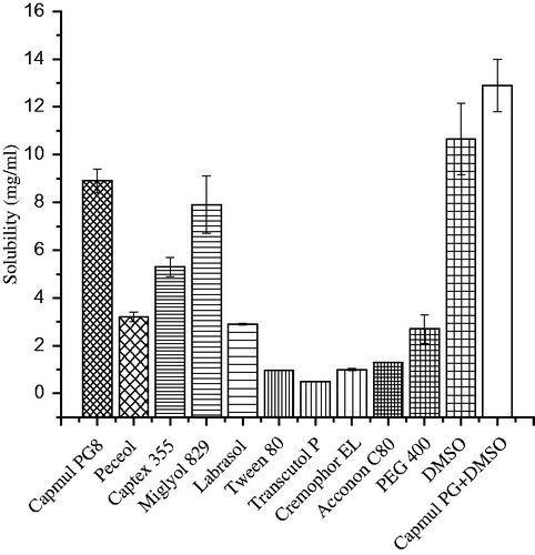
Figure 2. Photographs of the zone of inhibition: (A–C) the Petri plates revealing control, capmul PG8 (CPG8) and capmul MCM C8 (C8), respectively against C. albicans. (D–F) the Petri plates showing control, CPG8 and C-8, respectively against A. niger. The data represents the mean ± SD, (n = 3).
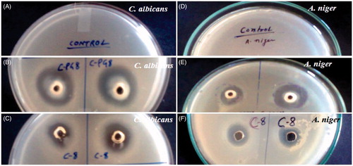
Table 1. Antifungal activities (zone of inhibition) against C. albicans and A. niger.
Figure 3. Pseudo-ternary phase diagrams of the optimized nanoemulsions delineated in different combinations of Smix as (A) and (B) reveal Smix ratio of 1:2 and 1:3, respectively.
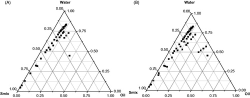
Table 2. Composition and characterization of developed nanoemulsion with their size, zeta potential, polydispersity index and ZOI (zone of inhibition) values.
Figure 4. Particle size, zeta potential and morphological studies of NE6: (A) particle size (nm) analysis, (B) zeta potential (mV), (C) transmission electron micrograph (TEM) of blank nanoemulsion and (D) TEM of drug-loaded nanoemulsion (AmB-NE6).
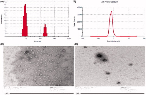
Figure 5. In vitro cumulative amount of Amphotericin B release through a dialysis membrane.
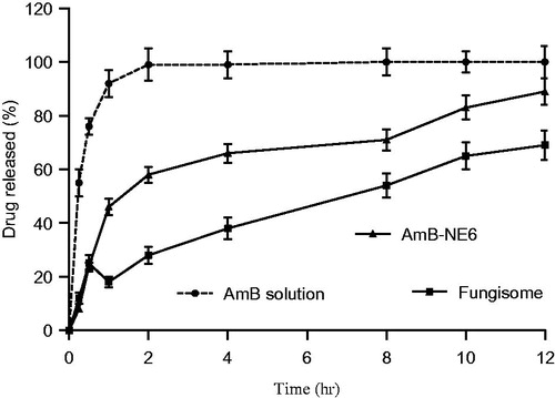
Figure 6. The cumulative amount of Amphotericin B across the albino rat skin using various formulations.
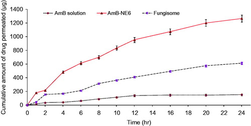
Table 3. Ex vivo permeation profile of different Amphotericin B formulations across the albino rat skin after 24 h of study.
Figure 7. The amount of drug deposited into the rat skin after ex vivo permeation study of various formulations and compared against commercial product Fungisome® (0.01% w/w).
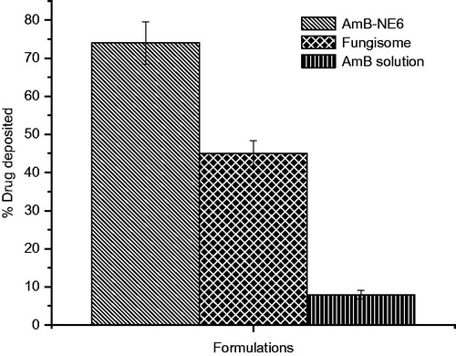
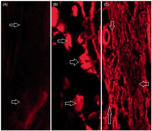
Figure 8. CLSM representative images depicting the penetration and distribution of Rhodamine-123 (Rh123) across albino rat skin after treatment: (A) Control Rh123 dye, (B) NE-Rh123 and (C) AmB-NE Rh123 after being applied for 8 h.