Figures & data
Figure 1. Arterial oxygen tension (PaO2) (Panel A), carbon dioxide tension (PaCO2) (Panel B) and pH (Panel C) versus time, plotted for pigs with arterial oxygen saturation (SaO2) > 90% pre-treatment (Hsat-Group, upper panels) and pigs with SaO2 < 90% pre-treatment (Lsat-Group, lower panels). Data shown for: air-breathing pre-shunt, during shunt application without and with (subsequent) oxygen breathing, and air-breathing at end of experiments. Number of animals in each time period shown in Panel A. Time and duration of infusions of dodecafluoropentane emulsion (DDFPe) marked in each panel. Values are means±SEM and P-values are shown for data of each time period marked by dashed lines compared with data from the last 10 min before DDFPe treatment.
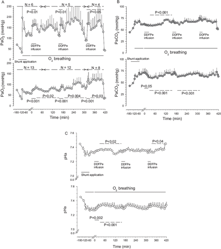
Figure 2. Oxygen tensions (PmO2) (Panel A) and carbon dioxide (PmCO2) (Panel B) tensions versus time in muscle tissue plotted in Hsat-Group and Lsat-Group pigs (for further explanations see legend).
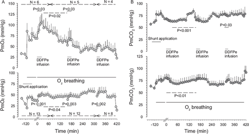
Figure 3. Central venous pressure (Panel A) and right ventricular pressure (Panel B) versus time in Hsat-Group and Lsat-Group pigs (for further explanations see legend).
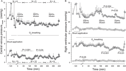
Figure 4. Average pulmonary artery pressure versus time in Hsat-Group and Lsat-Group pigs combined during air breathing (control) before and after shunt application and subsequent oxygen breathing and 30 min after the first infusion of DDFPe. All values are given as means±SEM (for further explanations see legend).
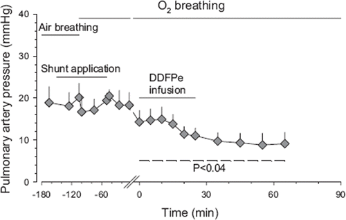
Table 1. Arterial and venous oxygen tensions and saturations in pigs during oxygen breathing after shunt application, before and during DDFPe treatment
Figure 5. This figure is based on theoretical calculations (see above) illustrating the effect, during oxygen breathing, of surface tension on the dose-response curve of DDFP emulsions in the treatment of hypoxia resulting from a 60% right to left shunt.
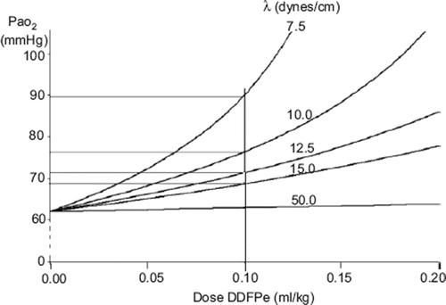
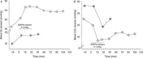
Figure 6. Panel A: arterial oxygen tension (PaO2), light grey circles and, venous oxygen tensions (PvO2), dark grey circles versus time in air-breathing pig with nosocomial pneumonia before, during and after infusion of 0.1 ml/kg (body weight) of DDFPe. Panel B: arterial carbon dioxide (PaCO2), light grey diamonds, and venous carbon dioxide (PvCO2) tensions, dark grey diamonds, in the same pig.