Figures & data
Figure 1. Fibrinogen isolation consists of three steps: separation of cruor and serum, fibrinogen precipitation, and preparation of the fibrinogen pellet by centrifugation.
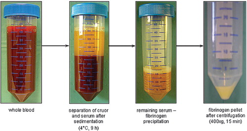
Figure 2. Schema for the first test series.
Figure 3. Schema for the second test series.
Figure 4. Impact of different temperatures for the gravity-sedimentation step: fibrinogen yield (wet weight in mg) was significantly higher using −20°C than using −86°C or −196°C (data represent the average + SD).
Figure 5. Fibrinogen yield (wet weight in mg) after the first isolation step using centrifugation at 200×g for 10 minutes vs. sedimentation at 4°C for 9 hours; 241 mg fibrinogen (wet weight) could be isolated out of 20 ml of patient whole blood using centrifugation and 309 mg fibrinogen (wet weight) using the sedimentation procedure (data represent the average + SD).
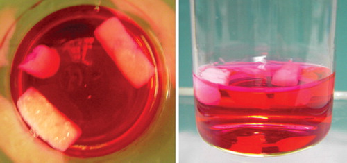
Figure 6. Fibrin glue constructs maintain form stability, size, and consistency after two weeks under cell culture conditions in DMEM (5% CO2, 37°C).