Figures & data
Figure 1. Mean arterial pressure () and mean pulmonary artery pressure () decrease during the first 15 minutes post-injury, when no resuscitation is given. Following resuscitation, MAP was restored to baseline at 120 minutes in the HBOC group, at 45 minutes in the HBOC+rFVIIa 90 μg/kg group, and at 105 minutes in the HBOC+rFVIIa 180 μg/kg group, whereas it did not return to baseline until 195 minutes in the HBOC+rFVIIa 360 μg/kg animals (p=0.0424). Mean pulmonary artery pressure (MPAP) was similar between groups throughout the pre-hospital phase (p=0.49; ). No groups demonstrated pulmonary hypertension; MPAP surpassed baseline only mildly in all groups.
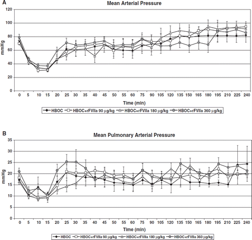
Figure 2. Heart rate in the HBOC+rFVIIa 90 μg/kg group was higher at the beginning of resuscitation and continued to increase until 120 minutes, at which point tachycardia began to resolve; in the other groups, tachycardia began to resolve by 60 minutes (). Cardiac Index (CI) was lowest in HBOC only pigs throughout the pre-hospital phase, and never returned to baseline. CI was similar between other treatment groups and was restored to baseline faster at 135 minutes in the HBOC+rFVIIa 90 μg/kg group than in the HBOC+rFVIIa 180 μg/kg (195 min.), and in the HBOC+rFVIIa 360 μg/kg groups (by the end of the pre-hospital phase (p<0.0001), ).
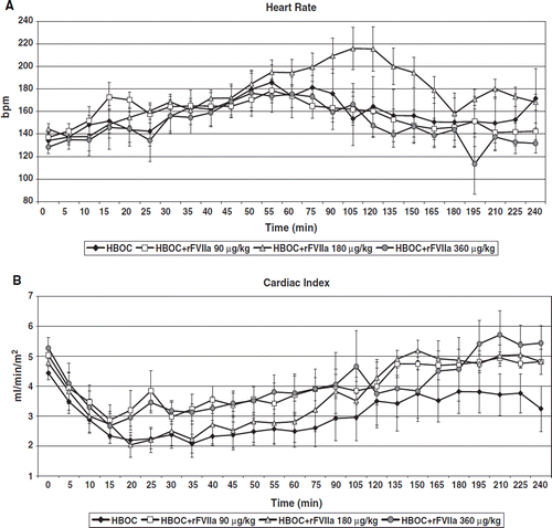
Table 1. Pre-hospital blood loss, fluid requirements, urine output and rFVIIa doses.
Figure 3. Pre-hospital fluid requirements were not statistically different between groups, but there was a trend towards reduced resuscitation requirement in the HBOC-201 only group compared to rFVIIa groups.
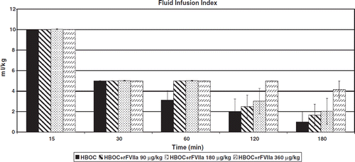
Figure 4. Lactate () increased in all groups in response to hemorrhage and remained above baseline in all groups with the exception of the HBOC+rFVIIa 90 μg/kg group (p=0.01). It increased substantially in the HBOC+rFVIIa 360 μg/kg group at 150 and 180 minutes (p = 0.05). Transcutaneous oxygen saturation (tcpO2) was significantly higher over time in the animals receiving 90 or 180 μg/kg rFVIIa compared to 360 μg/kg or HBOC alone (p<0.0001; ).
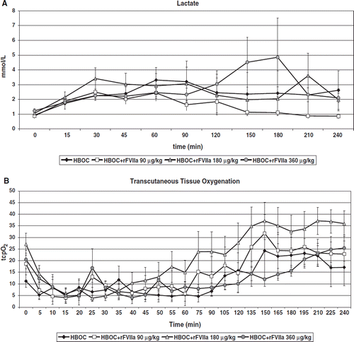
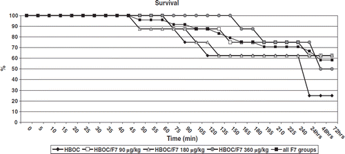
Figure 5. Survival to simulated hospital arrival (240 minutes) 5/8 (62.5%), 6/8 (75%), 5/8 (62.5%), 6/8 (75%) and 17/24 (70.8%) in HBOC, HBOC+rFVIIa 90 μg/kg, HBOC+rFVIIa 180 μg/kg, HBOC+rFVIIa 360 μg/kg and all rFVIIa groups-combined groups. Respective survival rates to 72 hours were 2/8 (25%), 5/8 (62.5%), 5/8 (62.5%), 4/8 (50%) and 14/24 (70.8%), p = 0.09.