Figures & data
Figure 1. Illustration of surgical precedures. A: Proximal median/ulnar nerve segment was served as father nerve to repair the distal nerve stump (Dor-Dor, Group 2, 3); B: Serving as a donor nerve, the proximal 1/2 median nerve was fixed to the distal stumps of 1/2 median and ulnar nerve simultaneously, using biodegegradable chitin conduits with a gap of 1 mm. (1/2Dor-1/2Dor+Rec, Group 4).
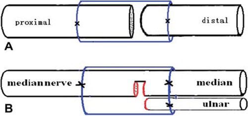
Table 1. Comparison of myelinated axon numbers for all groups.
Figure 2. Histological sections through the reconstructed nerves 4 months after surgery. Proximal part of the median nerve (A) and ulnar nerve (B). Distal stump of transected median (C, Dor-Dor, Group 2) or ulnar (D, Dor-Dor Group 3) nerve neurorrhaphy; Distal regenerated median (E) and ulnar (F) stump of the group (1/2Dor-1/2Dor+Rec Group 4) in which half proximal median proximal median nerve was served as donor nerve to reconstruct the distal 1/2 median and ulnar nerve stump. (G) Distal stump of the reconstructed median nerve. Both regenerated fibers and original intact fibers could be observed.
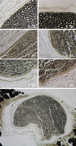
Figure 3. Nerve conduction velocities of rabbit regenerative median and ulnar nerve. Data were collected 4 months after reconstructive surgery. Electrophysiological recording was performed on the distal part of the median or ulnar nerve. There is no significant difference among the transected median/ulnar nerve neurorrhaphy groups (Dor-Dor, group 2, 3) and the group (1/2Dor-1/2Dor+Rec, group 4) in which proximal half median nerve served as donor nerve to reconstruct the distal median and ulnar nerve stump.
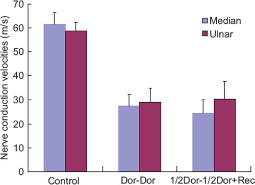
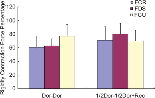
Figure 4. The tetanic muscle force in the flexor carpi radialis muscle (FCR), flexor digitorum superficialis (FDS) and the flexor carpi ulnaris (FCU). It showed that the ipsilateral side had values of 61 ± 16% 63 ± 10% 77 ± 17% of the contralateral side, after the distal ulnar and median nerves were repaired by their own proximal stem (Dor-Dor, Group 2, 3). And the values were 71 ± 20% 80 ± 16% 70 ± 16% in the group where distal ulnar and 1/2 median nerves were reconstructed by the proximal half median nerve (1/2 Dor-1/2Dor+Rec, Group 4).