793
Views
18
CrossRef citations to date
0
Altmetric
Research Article

Fluid Resuscitation of Hemorrhagic Shock with Hemoglobin Vesicles in Beagle Dogs: Pilot Study

, , , , , & show all
Pages 179-195 | Published online: 31 Jan 2012

Figures & data

Figure 1a. Effects of Lactated Ringer's solution (LR), human serum albumin (HSA), autologous shed blood (ASB), and hemoglobin vesicles (HbV) on heart rate (HR) in anesthetized dogs subjected to 50% exsanguination. There were no significant differences between the HbV group and the other groups. (○: LR group; □: HSA group, × : ASB group; △: HbV group).
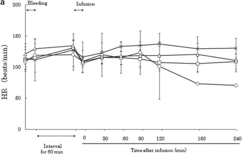
Figure 1a. Effects of Lactated Ringer's solution (LR), human serum albumin (HSA), autologous shed blood (ASB), and hemoglobin vesicles (HbV) on heart rate (HR) in anesthetized dogs subjected to 50% exsanguination. There were no significant differences between the HbV group and the other groups. (○: LR group; □: HSA group, × : ASB group; △: HbV group).

Figure 1b. Effects of Lactated Ringer's solution (LR), human serum albumin (HSA), autologous shed blood (ASB), and hemoglobin vesicles (HbV) on MAP (mean arterial pressure) in anesthetized dogs subjected to 50% exsanguination. There were significant differences between the LR and HbV groups 30 minutes after resuscitation. (*) (○: LR group; : HSA group; × : ASB group; △: HbV group).
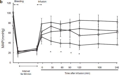
Figure 1b. Effects of Lactated Ringer's solution (LR), human serum albumin (HSA), autologous shed blood (ASB), and hemoglobin vesicles (HbV) on MAP (mean arterial pressure) in anesthetized dogs subjected to 50% exsanguination. There were significant differences between the LR and HbV groups 30 minutes after resuscitation. (*) (○: LR group; : HSA group; × : ASB group; △: HbV group).

Figure 1c. Effects of Lactated Ringer's solution (LR), human serum albumin (HSA), autologous shed blood (ASB), and hemoglobin vesicles (HbV) on mean pulmonary arterial pressure (MPAP) in anesthetized dogs subjected to 50% exsanguination. There were no significant differences between the HbV group and the other groups. (○: LR group; □: HSA group; × : ASB group; △: HbV group).
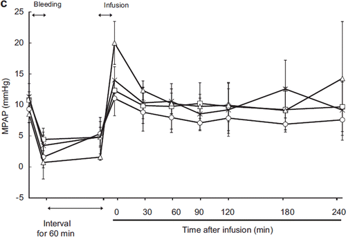
Figure 1c. Effects of Lactated Ringer's solution (LR), human serum albumin (HSA), autologous shed blood (ASB), and hemoglobin vesicles (HbV) on mean pulmonary arterial pressure (MPAP) in anesthetized dogs subjected to 50% exsanguination. There were no significant differences between the HbV group and the other groups. (○: LR group; □: HSA group; × : ASB group; △: HbV group).

Figure 1d. Effects of Lactated Ringer's solution (LR), human serum albumin (HSA), autologous shed blood (ASB), and hemoglobin vesicles (HbV) on pulmonary capillary wedge pressure (PCWP) in anesthetized dogs subjected to 50% exsanguination. Value is expressed as a ratio to that of the baseline. There were significant differences between the LR group and the HSA, ASB, and HbV groups (η) and between the HSA and HbV and ASB groups (*). (○: LR group; □: HSA group; × : ASB group; △: HbV group).
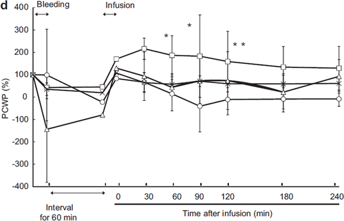
Figure 1d. Effects of Lactated Ringer's solution (LR), human serum albumin (HSA), autologous shed blood (ASB), and hemoglobin vesicles (HbV) on pulmonary capillary wedge pressure (PCWP) in anesthetized dogs subjected to 50% exsanguination. Value is expressed as a ratio to that of the baseline. There were significant differences between the LR group and the HSA, ASB, and HbV groups (η) and between the HSA and HbV and ASB groups (*). (○: LR group; □: HSA group; × : ASB group; △: HbV group).

Figure 2a. Effects of Lactated Ringer's solution (LR), human serum albumin (HSA), autologous shed blood (ASB), and hemoglobin vesicles (HbV) on cardiac output (CO) in anesthetized dogs subjected to 50% exsanguination. There were significant differences between the LR and HSA groups (η) and between the HSA and HbV groups (*). ( : LR group; □: HSA group; × : ASB group; △: HbV group).
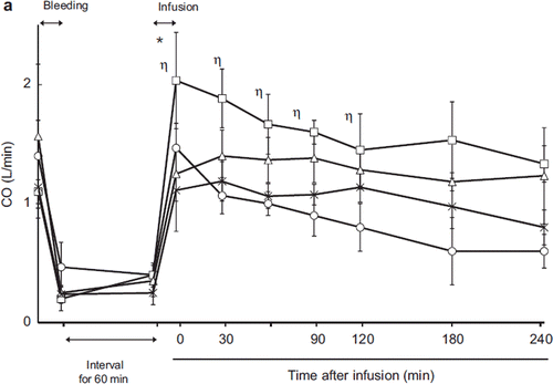
Figure 2a. Effects of Lactated Ringer's solution (LR), human serum albumin (HSA), autologous shed blood (ASB), and hemoglobin vesicles (HbV) on cardiac output (CO) in anesthetized dogs subjected to 50% exsanguination. There were significant differences between the LR and HSA groups (η) and between the HSA and HbV groups (*). ( : LR group; □: HSA group; × : ASB group; △: HbV group).

Figure 2b. Effects of Lactated Ringer's solution (LR), human serum albumin (HSA), autologous shed blood (ASB), and hemoglobin vesicles (HbV) on systemic vascular resistance (SVR) ratio in anesthetized dogs subjected to 50% exsanguination. There were significant differences between the HSA and HbV groups after resuscitation. (○; LR group, □; HSA group, × ; ASB group, △; HbV group ).
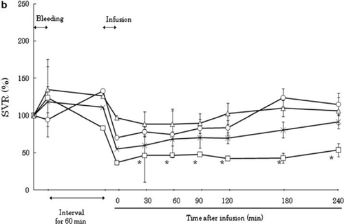
Figure 2b. Effects of Lactated Ringer's solution (LR), human serum albumin (HSA), autologous shed blood (ASB), and hemoglobin vesicles (HbV) on systemic vascular resistance (SVR) ratio in anesthetized dogs subjected to 50% exsanguination. There were significant differences between the HSA and HbV groups after resuscitation. (○; LR group, □; HSA group, × ; ASB group, △; HbV group ).

Figure 3a. Effects of Lactated Ringer's solution (LR), human serum albumin (HSA), autologous shed blood (ASB), and hemoglobin vesicles (HbV) on PaO2 in anesthetized dogs subjected to 50% exsanguination. There were no significant differences between the HbV group and the other groups. (○: LR group; □: HSA group; × : ASB group; △: HbV group).
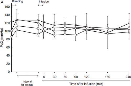
Figure 3a. Effects of Lactated Ringer's solution (LR), human serum albumin (HSA), autologous shed blood (ASB), and hemoglobin vesicles (HbV) on PaO2 in anesthetized dogs subjected to 50% exsanguination. There were no significant differences between the HbV group and the other groups. (○: LR group; □: HSA group; × : ASB group; △: HbV group).

Figure 3b. Effects of Lactated Ringer's solution (LR), human serum albumin (HSA), autologous shed blood (ASB), and hemoglobin vesicles (HbV) on PvO2 in anesthetized dogs subjected to 50% exsanguination. There were significant differences between the LR group and the other groups 30 minutes after resuscitation (*). (○: LR group; □: HSA group; × : ASB group; △: HbV group).
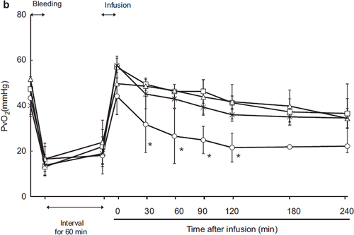
Figure 3b. Effects of Lactated Ringer's solution (LR), human serum albumin (HSA), autologous shed blood (ASB), and hemoglobin vesicles (HbV) on PvO2 in anesthetized dogs subjected to 50% exsanguination. There were significant differences between the LR group and the other groups 30 minutes after resuscitation (*). (○: LR group; □: HSA group; × : ASB group; △: HbV group).

Figure 4a. Effects of Lactated Ringer's solution (LR), human serum albumin (HSA), autologous shed blood (ASB), and hemoglobin vesicles (HbV) on PtO2 of the renal cortex in anesthetized dogs subjected to 50% exsanguination. There were no significant differences between the HbV group and the other groups. (○: LR group; □: HSA group; × : ASB group; △: HbV group).
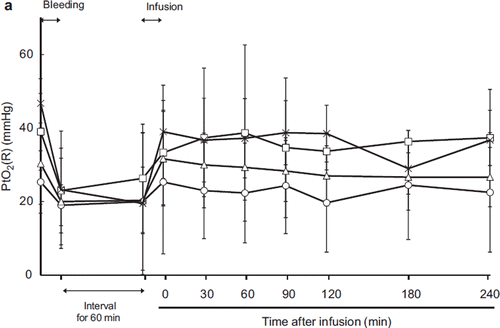
Figure 4a. Effects of Lactated Ringer's solution (LR), human serum albumin (HSA), autologous shed blood (ASB), and hemoglobin vesicles (HbV) on PtO2 of the renal cortex in anesthetized dogs subjected to 50% exsanguination. There were no significant differences between the HbV group and the other groups. (○: LR group; □: HSA group; × : ASB group; △: HbV group).
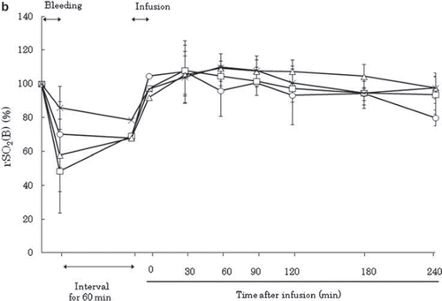
Figure 4b. Effects of Lactated Ringer's solution (LR), human serum albumin (HSA), autologous shed blood (ASB), and hemoglobin vesicles (HbV) on regional oxygen saturation of the brain (rSO2 (B)) in anesthetized dogs subjected to 50% exsanguination. The value was expressed as a ratio to the baseline. There were no significant differences between the HbV group and the other groups. (○: LR group; □: HSA group; × : ASB group; △: HbV group).

Figure 4b. Effects of Lactated Ringer's solution (LR), human serum albumin (HSA), autologous shed blood (ASB), and hemoglobin vesicles (HbV) on regional oxygen saturation of the brain (rSO2 (B)) in anesthetized dogs subjected to 50% exsanguination. The value was expressed as a ratio to the baseline. There were no significant differences between the HbV group and the other groups. (○: LR group; □: HSA group; × : ASB group; △: HbV group).

Figure 5a. Effects of Lactated Ringer's solution (LR), human serum albumin (HSA), autologous shed blood (ASB), and hemoglobin vesicles (HbV) on hematocrit (Ht) in anesthetized dogs subjected to 50% exsanguination. There were no significant differences between the HbV group and the HSA and LR groups. (○: LR group; □: HSA group; × : ASB group; △: HbV group).
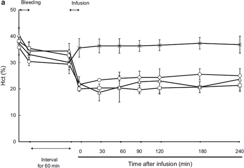
Figure 5a. Effects of Lactated Ringer's solution (LR), human serum albumin (HSA), autologous shed blood (ASB), and hemoglobin vesicles (HbV) on hematocrit (Ht) in anesthetized dogs subjected to 50% exsanguination. There were no significant differences between the HbV group and the HSA and LR groups. (○: LR group; □: HSA group; × : ASB group; △: HbV group).

Figure 5b. Contribution of HbV to the total hemoglobin concentration after administration of HbV. (Black bar: Hb derived from HbV; blank bar: Hb derived from RBC).
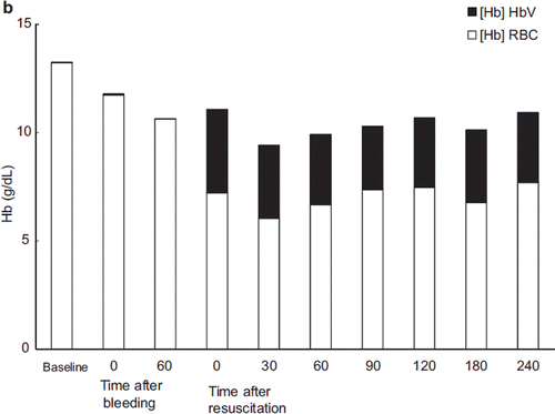
Figure 5b. Contribution of HbV to the total hemoglobin concentration after administration of HbV. (Black bar: Hb derived from HbV; blank bar: Hb derived from RBC).

Figure 6a. Effects of Lactated Ringer's solution (LR), human serum albumin (HSA), autologous shed blood (ASB), and hemoglobin vesicles (HbV) on pH in anesthetized dogs subjected to 50% exsanguination. There were no significant differences between the HbV group and the other groups. (○: LR group; □: HSA group; × : ASB group; △: HbV group).
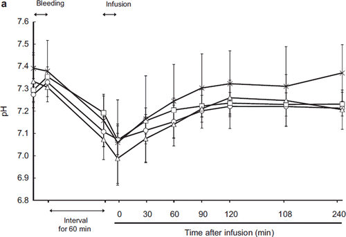
Figure 6a. Effects of Lactated Ringer's solution (LR), human serum albumin (HSA), autologous shed blood (ASB), and hemoglobin vesicles (HbV) on pH in anesthetized dogs subjected to 50% exsanguination. There were no significant differences between the HbV group and the other groups. (○: LR group; □: HSA group; × : ASB group; △: HbV group).

Figure 6b. Effects of Lactated Ringer's solution (LR), human serum albumin (HSA), autologous shed blood (ASB), and hemoglobin vesicles (HbV) on serum lactate level in anesthetized dogs subjected to 50% exsanguination. There were no significant differences between the HbV group and the other groups. ( ○: LR group; □: HSA group; × : ASB group; △: HbV group).
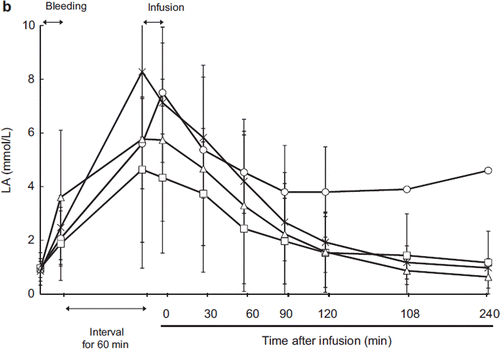
Figure 6b. Effects of Lactated Ringer's solution (LR), human serum albumin (HSA), autologous shed blood (ASB), and hemoglobin vesicles (HbV) on serum lactate level in anesthetized dogs subjected to 50% exsanguination. There were no significant differences between the HbV group and the other groups. ( ○: LR group; □: HSA group; × : ASB group; △: HbV group).

Figure 7. Proportion of MetHb in HbV hemoglobin. The concentration of methemoglobin gradually increased.
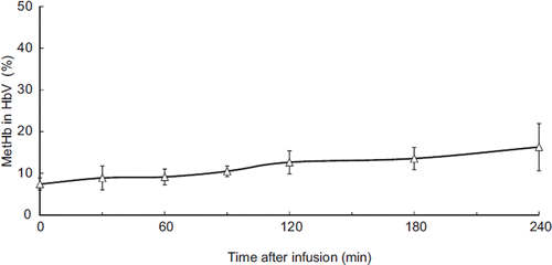
Figure 7. Proportion of MetHb in HbV hemoglobin. The concentration of methemoglobin gradually increased.

Figure 8a. Contribution of HbV to oxygen delivery. HbV delivered 33–26% of the tissue oxygen supply. (Black bar: oxygen delivered by HbV; blank bar: oxygen delivered by RBC).
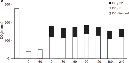
Figure 8a. Contribution of HbV to oxygen delivery. HbV delivered 33–26% of the tissue oxygen supply. (Black bar: oxygen delivered by HbV; blank bar: oxygen delivered by RBC).

Figure 8b. Contribution of HbV to oxygen consumed. HbV conveyed 25–20% of oxygen used. Black bar: calculated oxygen amount consumed from HbV; blank bar: calculated oxygen amount consumed from RBC.
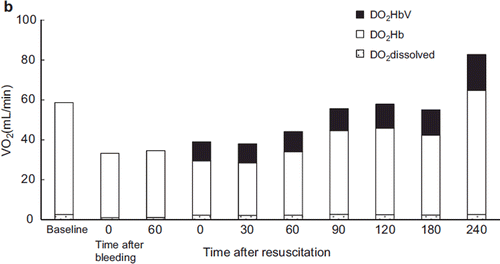
Figure 8b. Contribution of HbV to oxygen consumed. HbV conveyed 25–20% of oxygen used. Black bar: calculated oxygen amount consumed from HbV; blank bar: calculated oxygen amount consumed from RBC.

Table 1. Results of serum chemistry analysis.

Reprints and Corporate Permissions

Please note: Selecting permissions does not provide access to the full text of the article, please see our help page How do I view content?

To request a reprint or corporate permissions for this article, please click on the relevant link below:

Academic Permissions

Please note: Selecting permissions does not provide access to the full text of the article, please see our help page How do I view content?

Obtain permissions instantly via Rightslink by clicking on the button below:

If you are unable to obtain permissions via Rightslink, please complete and submit this Permissions form. For more information, please visit our Permissions help page.