Figures & data
Figure 1. Porcine oral mucosa models analyzed by histology by using HPS staining (a,b,c), and by immunolabeling of the protein keratin 13, the major differentiation marker of non-keratinized oral epithelium (d,e,f). Immunolabeling is shown in green, and cell nuclei in red (x200).
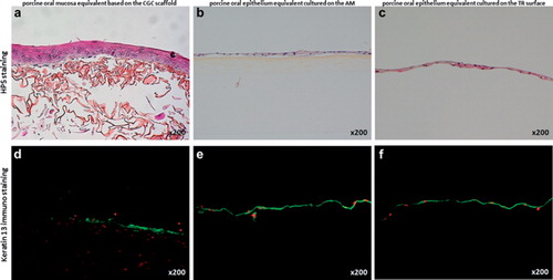
Figure 2. Transmission electron microscopic analysis of the porcine oral mucosa models. Numerous intercellular junctions such as desmosomes (D) were detected in the epithelium of the model based on the CGC scaffold (a), the epithelium cultured on the AM (b), and the reconstructed epithelial cell sheet (c). In the lamina propria equivalent of the CGC-based 3D model, transversal and longitudinal sections of the newly synthesized collagen fibers (C) by the active porcine fibroblasts were visible (d). Bars = 200 nm.