Figures & data
Figure 1. Schematic diagram of experimental animal models. (A): The common peroneal nerve was transected 5 mm below the sciatic nerve bifurcation point and the distal and proximal nerve stumps were bridged at the breakpoint using the conduit. Three months after the first surgery, the common peroneal and tibial nerves were transected 1 cm below the first breakpoint and the proximal common peroneal nerve was used as the donor to repair the distal tibial nerve after it was also transected. Schematic diagram of control group animal models (B): The control group animals were anesthetized and the sciatic nerve was exposed without surgery and the wound was closed. Three months after the first surgery, the common peroneal and tibial nerves were transected and the proximal common peroneal nerve and the distal tibial nerve were bridged using the conduit.
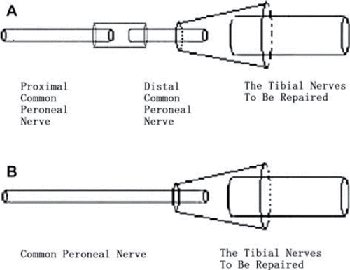
Figure 2. Images of the transverse section of the nerve following osmium tetroxide staining (X400): The normal tibial nerve (A); the normal peroneal nerve (B); the donor nerve of the second recovery process in the experimental group (C); the receptor nerve of the second recovery process in the experimental group (D); the donor nerve of the second recovery process in the control group (E); the receptor nerve of the second recovery process in the control group (F).
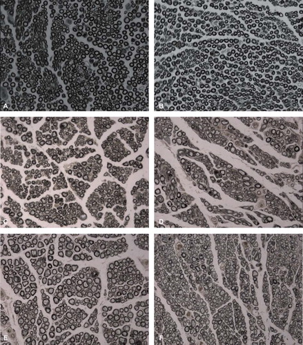
Table I. Number of the myelinated nerve fibers and RDP.
Figure 3. The waveform of normal gastrocnemius compound action potential (A); the waveform of gastrocnemius compound action potential in the experimental group (B); the waveform of gastrocnemius compound action potential in the control group (C).
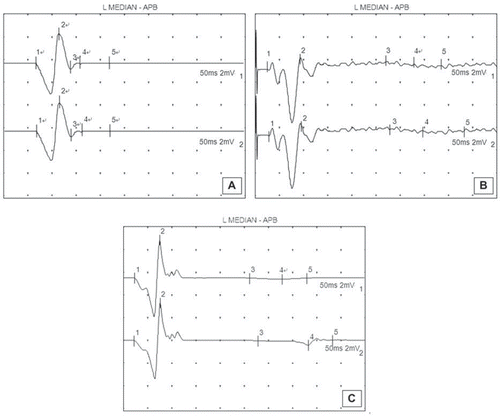
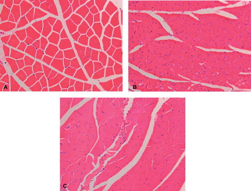
Figure 4. Images of the transverse section of the tibialis anterior muscle, following hematoxylin and eosin staining (X200), compared with the contralateral normal muscle (A). Different degrees of muscle atrophy were seen in the operated side of the experimental (B) and control groups (C).