Figures & data
Figure 1. SEM micrograph of CEPs-loaded chitosan microparticles.
Figure 2. Particle size distribution of (a) CEPs-loaded chitosan microparticles and (b) chitosan microparticles.
Figure 3. Release of CEPs from CEPs-loaded chitosan microparticles at pH 7.4 and 37茄C (data shown are the mean ± SD, n = 3).
Figure 4. SDS-PAGE (12 % gel) of CEPs from V. cholerae. Lanes: (1) CEPs as native antigen; (2) release of CEPs in PBS at 37茄C.
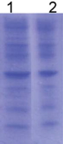
Figure 5. Secretion of IFN-γ form THP1 cell line stimulated with (a) CEPs loaded chitosan microparticle; (b) CEPs; and (c) control (n = 3).
Figure 6. Secretion of TNF α form THP1 cell line stimulated with (a) CEPs loaded chitosan microparticles; (b) CEPs; and (c) control (n = 3).
Figure 7. Secretion of IL-6 form THP1 cell line stimulated with (a) CEPs loaded chitosan microparticles; (b) CEPs; and (c) control (n = 3).