Figures & data
Table I. Primer sequences used in RT-PCR and qPCR assay.
Figure 1. Morphology and growth curves of BMMSCs (100×) at (A) P5, (B) P15, (C) P25, (D) P35, (E) P45, and at (F) passages 10, 25 and 45 for comparison. The cells expanded easily and exhibited fibroblast-like morphology. The growth curves of BMMSCs at different passages were S-shaped, and the PDTs of BMMSCs were 30.21, 31.78, 35.19 and 35.87 hr respectively.
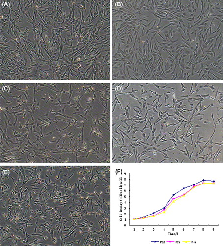
Figure 2. Surface antigen characteristics of BMMSCs at different passages. Immunoflourescence staining results showed that BMMSCs at different passages expressed antigens CD44, CD29, ICAM-1, CD71, CD73, CD90, CD105 and SSEA-4, but not antigens CD31 and CD34.
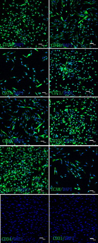
Figure 3. Detection of surface markers of BMMSCs via RT-PCR. RT-PCR assays showed that BMMSCs at different passages were positive for CD29, ICAM-1, SSEA-4, CD44, CD71 and negative for CD73, CD31 and CD34.
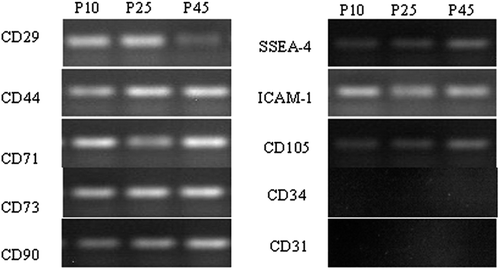
Figure 4. Osteogenic differentiation of BMMSCs. A: The control group. B: After incubation in osteogenic medium for 17 days, the cells metamorphosed from fusiform to tridimensional shapes and the nodules increased in number and size with prolonged inducing time. C: About 20 days later, the nodules were obviously observed following Alizarin Red staining. Cells cultured in complete medium were not influenced in morphology or stained by Alizarin Red (200×).
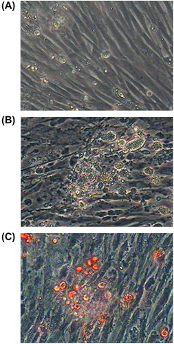
Figure 5. Detection of ALP concentration in inducer medium. The ALP concentration assay of medium showed that ALP concentration increased with induced time extension. Group C are significantly different (P < 0.05) than other group.

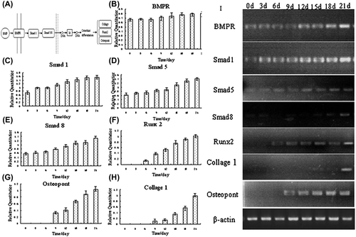
Figure 6. PCR analyses of BMP signals pathway members. A BMP regulation of osteoblast differentiation, ║ cell membrane, ║ karyotheca,Id inhibitor of DNA binding, dominant negative helix-loop-helix protein. B to H qPCR analyses of BMP signals pathway members. I RT-PCR analyses of BMP signals pathway members.