Figures & data
Figure 1. Preoperative diagnostics with an MRI scan of a patient with unspecific knee pain. The osteoid osteoma lesion was located in the postero-medial facet of the patella.
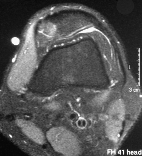
Figure 2. Preoperative CT scan of an osteoid osteoma lesion of the proximal medial tibia.
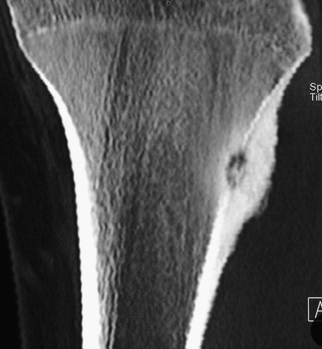
Figure 3. Intraoperative Iso-C3D® scan of the region of interest. The site was secondarily draped during the scanning procedure. Navigation tools are placed on an extra table.
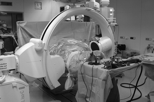
Figure 4. Intraoperative set-up of the navigation system related to the site and the Iso-C3D®.
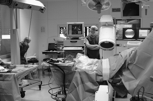
Figure 5. The DRB is fixed with two Schanz screws to the patella to achieve rotation stability during the drilling procedure. A navigated pointer was used to check the initial plausibility of the navigated tools.
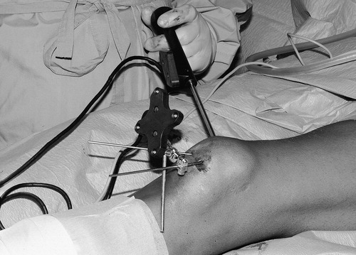
Figure 6. Fixation of the DRB in the proximal third of the tibia and plausibility check with the pointer.
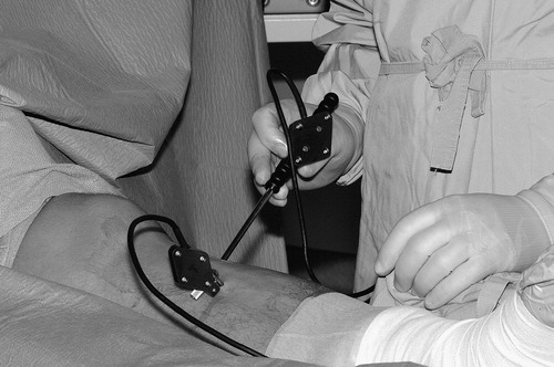
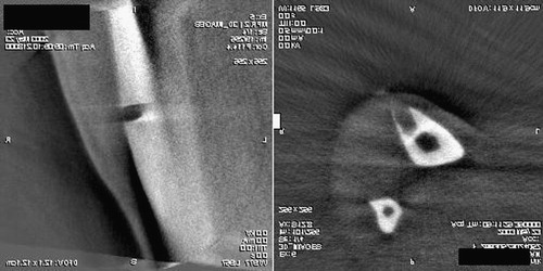
Figure 7. (a) Percutaneous navigated drilling of the proximal tibial lesion with a hollow drill. (b) Intraoperative navigation screen in three multiplanar reconstructions. The actual position of the drill (green) is shown in reference to the good visible lesion on the tibia shaft. An aiming device helps to drill along the planned trajectories (red) and shows the distance from the start point. [Color version available online]
![Figure 7. (a) Percutaneous navigated drilling of the proximal tibial lesion with a hollow drill. (b) Intraoperative navigation screen in three multiplanar reconstructions. The actual position of the drill (green) is shown in reference to the good visible lesion on the tibia shaft. An aiming device helps to drill along the planned trajectories (red) and shows the distance from the start point. [Color version available online]](/cms/asset/20507929-0430-4d3f-ad4a-7235e879f0d4/icsu_a_122969_f0007_b.jpg)
Figure 8. A conventional postoperative CT scan done 5 weeks after surgery on the patient with the distal tibial shaft lesion. The scan is congruent with the direct intraoperative images of the Iso-C3D® control scan ().
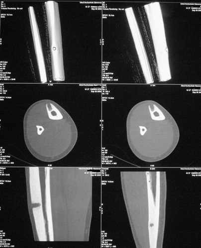
Figure 9. Intraoperative Iso-C3D® control scan after navigated resection of the lesion at the distal tibia.