Figures & data
Figure 1. Left: Ethmoidal cells Citation[18] are often the source of the inflamed tissue. Right: The preoperative CT scan shows polyps in the right sinus sphenoidalis.
![Figure 1. Left: Ethmoidal cells Citation[18] are often the source of the inflamed tissue. Right: The preoperative CT scan shows polyps in the right sinus sphenoidalis.](/cms/asset/22a0f418-5ab8-45a0-a837-ee1b575c0c4b/icsu_a_175041_f0001_b.gif)
Figure 2. The shaver is a motor-driven instrument used for the removal of inflamed tissue.
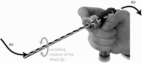
Figure 3. The workflow of the system comprises three steps: preparation, preoperative surgical planning, and surgical intervention.
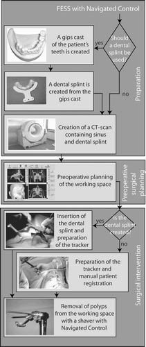
Figure 4. The system contains the following hardware components: navigation system, navigated control electronics, shaver control unit, foot pedal, shaver, and patient tracker.
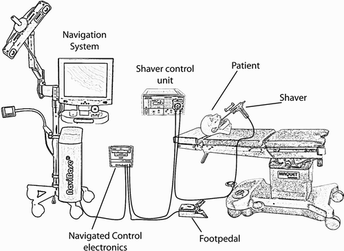
Figure 5. The preoperative planning module allows visualization of the axial, coronal and sagittal views of the 3D surface and enables the definition of a working space for the shaver.
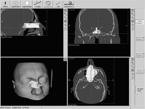
Figure 6. An optical tracking array can be easily connected to the shaver.
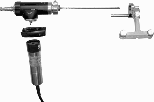
Figure 7. Intraoperatively, the surgeon sees the position of the shaver relative to the preoperative CT data and to the preoperatively defined working space.
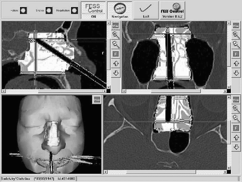
Figure 8. The working space is modeled as a voxel volume. The size of the voxels in the figure is greater than the size of real voxels compared to the dimensions of the shaver (the shaver has a diameter of 4 mm; the typical height of the voxels is 0.5 mm).
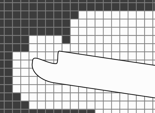
Figure 9. The navigated control electronics is a core component that connects all the other components and controls the shaver.
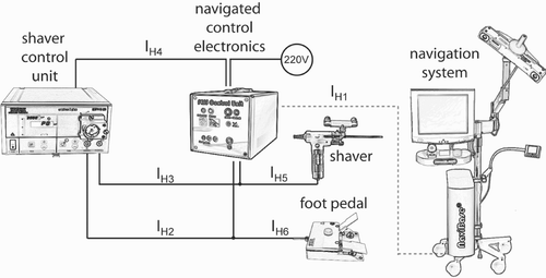
Figure 10. Different specimens can be placed in the technical model. Reproducible placement is realized with magnetic clips familiar from dental cast models.
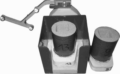
Figure 11. The test subject had to remove the foam manually by visually inspecting an existing template.
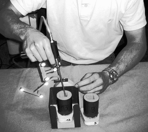
Figure 12. A rectangular cavity was defined in the planning.
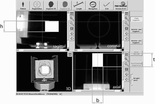
Table 1 The time for tissue removal was measured for 15 specimens. The time required with navigated control is equal to that for manual tissue removal, and less than that for removal with a navigation system.
Figure 13. The cavity was measured with a precise coordinate measuring machine. The coordinate transformation was achieved using a metal plate, whose coordinates relative to the dental splint were known.
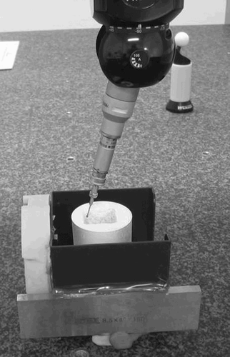
Figure 14. The sampled points recorded by the coordinate measuring machine are shown relative to the planned cavity.
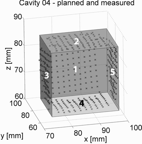
Table II. For the precision measurement, approximately 90 points were recorded on the 5 walls of the 5 specimens. The distance between these points and the planned cavity was calculated, and the mean, maximal and minimal distances, as well as the standard deviation, are displayed in this table.
Figure 15. The surgeons planned 7 patients; only the planning for the first patient took longer than 5 min.
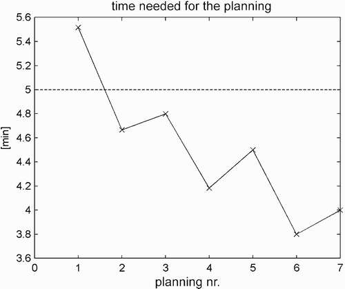
Table III. Planning times for the seven trials. Only the first planning time exceeded 5 minutes.
Figure 16. The system was evaluated in a clinical trial at the University of Leipzig, Germany.