Figures & data
Figure 1. Relationships between the sensor and CT coordinate systems.
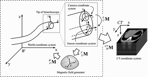
Figure 2. Flowchart of the proposed method.
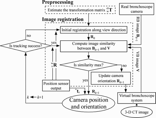
Figure 3. Example of the bronchoscope phantom model. [Color version available online]
![Figure 3. Example of the bronchoscope phantom model. [Color version available online]](/cms/asset/3444830c-d4d3-4d46-a512-474e2b1ea2f0/icsu_a_175078_f0003_b.jpg)
Figure 4. Layout of inserted acrylic pipes. The phantom was fixed in epoxy resin inside a plastic box, and 24 acrylic pipes were inserted.
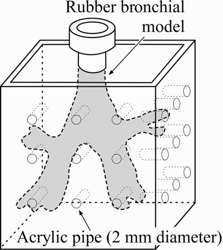
Figure 5. Experimental environment. [Color version available online]
![Figure 5. Experimental environment. [Color version available online]](/cms/asset/3fa8058a-917a-425f-9113-69cbbcdf2592/icsu_a_175078_f0005_b.jpg)
Figure 6. Results for FRE and RE obtained by changing the number of measured positions for computing transformation matrix .
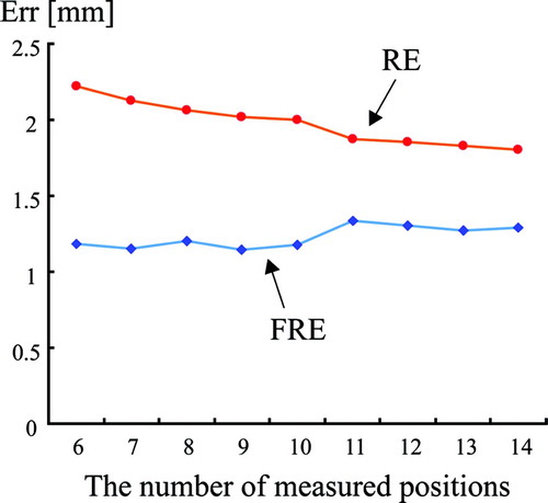
Table I. Standard deviations of the sensor's positional output. The sensor was placed 200 mm from the magnetic field generator, and the positional output was measured 600 times every 0.1 seconds for each of two positions: (i) with the sensor outside the bronchoscope; (ii) with the sensor inserted into the bronchoscope.
Figure 7. Tracking results of the proposed method. The left column shows scenes from the experiment; the right column shows the corresponding tracked results. [Color version available online]
![Figure 7. Tracking results of the proposed method. The left column shows scenes from the experiment; the right column shows the corresponding tracked results. [Color version available online]](/cms/asset/3d97c032-2d30-400b-95d6-d559d82fc225/icsu_a_175078_f0007_b.jpg)
Figure 8. Tracking results of the proposed method at several positions of the bronchus (trachea, left main bronchus, and intermediate bronchus). [Color version available online]
![Figure 8. Tracking results of the proposed method at several positions of the bronchus (trachea, left main bronchus, and intermediate bronchus). [Color version available online]](/cms/asset/7a4a2b8a-b435-44e3-b459-2f08520dc686/icsu_a_175078_f0008_b.jpg)
Figure 9. Tracking results of the method using only sensor output. [Color version available online]
![Figure 9. Tracking results of the method using only sensor output. [Color version available online]](/cms/asset/53cc6c75-8051-477b-8703-694f6bad58bf/icsu_a_175078_f0009_b.jpg)