Figures & data
Figure 1. A conceptual overview of the brachytherapy procedure
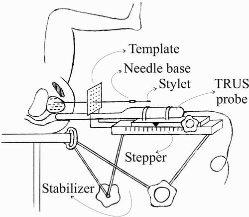
Figure 2. The observed prostate shift during needle insertion.
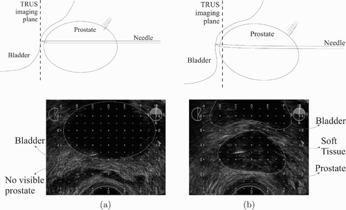
Figure 3. (a) Prostate surface obtained by Nuages, and (b) the axially symmetric 3D needle-mesh design.
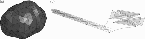
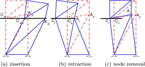
Figure 4. The simulation step (a) before and (b) after a new element penetration, where the tissue is remeshed using (c) node repositioning and (d) node addition techniques.
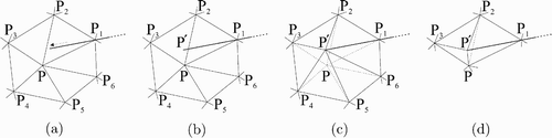
Figure 5. The flowchart of a typical iteration during 3D needle-tissue interaction simulation.
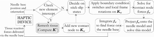
Figure 6. Computation time of node repositioning for varying mesh size (a) and node valence (b).
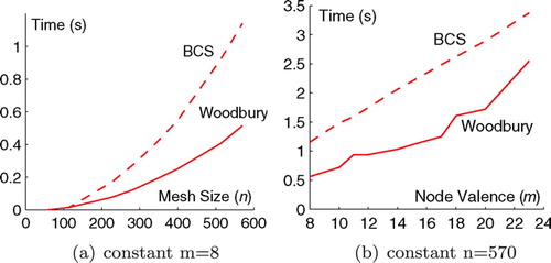
Figure 7. The tissue mesh prior to deformation (a), and while the needle is inserted (b, c), moved upwards (d), and retracted (e, f).
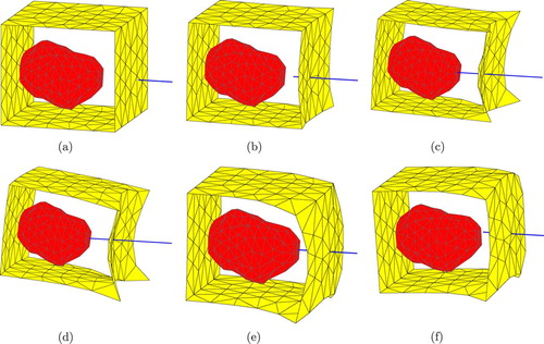
Figure 8. Axial, sagittal, and coronal forces on the needle base, and the sagittal torque.
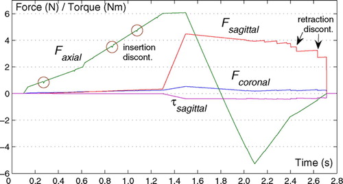
Figure 9. Tissue mesh (a) before and (b) after remeshing (note that does not end up at the intended position Pd without iterative optimization).
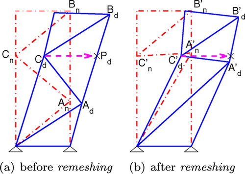
Figure 10. The torque accumulated in the tissue between (a) the insertion and (b) the retraction can be observed in (c) when the tip node slips off.