Figures & data
Figure 1. A customized prototype of the navigated parallel drill guide (PDG), equipped with a stable, integrated marker array. The reference markers can easily be adjusted for bilateral use.
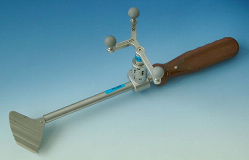
Figure 2. Visualization of the parallel drill guide displayed in the software. An aiming device and parallel screw planning allow for exact parallel screw placement within the radiographic landmarks. This trial was done on a plastic bone model. The upper right “AutoPilot” view visualizes the aiming of the actual drill or PDG (green broken line) according to the planned trajectory (red line), and shows discrepancies by deviations of the red dot from the center.
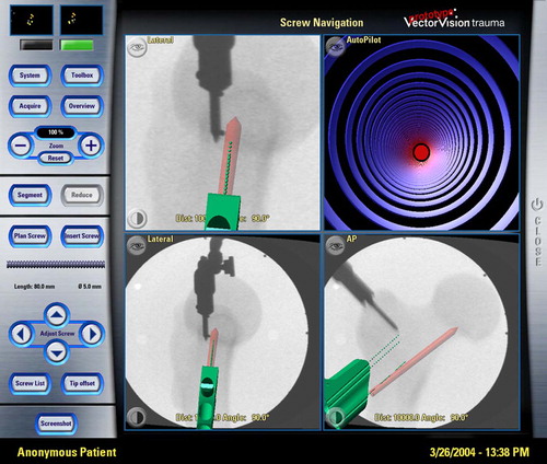
Figure 3. Navigation screen during the use of the navigated PDG on the cadaver trial. The three X-ray-based images show in axial and AP views the initially planned trajectory of the first caudal screw (red line). The two green lines visualize the potential aiming device, including the projected position of the caudal (green broken line) and cranial (green dotted line) drill canals through the PDG. The navigated drill bit positioned in the cranial screw canal is shown in brown with a larger diameter and displayed in combination with the PDG.
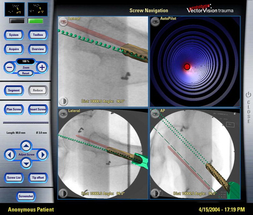
Figure 4. Combination of the navigated PDG with drill sleeves and the navigated drill bit in a cadaver trial during navigated fixation; both the navigated drill and the parallel drill guide (PDG) are displayed on the screen. The reference array on the bone is placed 20 cm distal to the surgical field.
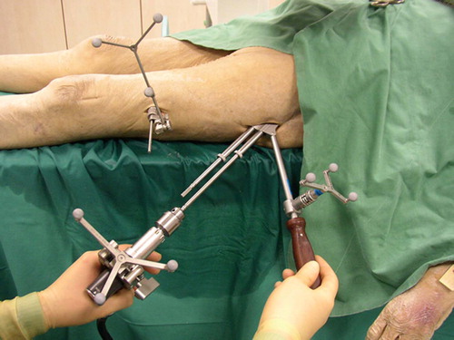
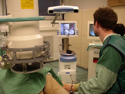
Figure 5. A dedicated set-up of all necessary systems facilitates cost-effective computer-assisted navigation. The set-up varies from that used with the conventional technique.