Figures & data
Figure 1. (a) Experimental system showing the positioning table to provide 6-DOF positioning and orientation of the magnetic sensor used to track the virtual tumor. (b) The modified RF scalpel containing a magnetic sensor. (c) Visual interface showing the disk inclined at 30° with the virtual scalpel showing the axial view of the task (top viewport) and coronal view (bottom viewport). (d) The navigation compass. [Color version available online.]
![Figure 1. (a) Experimental system showing the positioning table to provide 6-DOF positioning and orientation of the magnetic sensor used to track the virtual tumor. (b) The modified RF scalpel containing a magnetic sensor. (c) Visual interface showing the disk inclined at 30° with the virtual scalpel showing the axial view of the task (top viewport) and coronal view (bottom viewport). (d) The navigation compass. [Color version available online.]](/cms/asset/ba163c0b-39ef-4ca8-b663-1020d19ed530/icsu_a_225275_f0001_b.gif)
Figure 2. Typical experiment with volunteer accomplishing a “cutting” task on the virtual tumor generated with the setup shown in . Reproduced with kind permission of Springer Science and Business Media. [Color version available online.]
![Figure 2. Typical experiment with volunteer accomplishing a “cutting” task on the virtual tumor generated with the setup shown in Figure 1a. Reproduced with kind permission of Springer Science and Business Media. [Color version available online.]](/cms/asset/4c43880d-97a4-4f4f-87db-f14736e0a400/icsu_a_225275_f0002_b.gif)
Table I. Participant demographics.
Figure 3. (a) Positional error as a function of time for completion of experiment 18, including the total time, RMS error, and fraction of distance measurements taken inside the tumor. (b) The corresponding surgical path traced out by one volunteer in this experiment. [Color version available online.]
![Figure 3. (a) Positional error as a function of time for completion of experiment 18, including the total time, RMS error, and fraction of distance measurements taken inside the tumor. (b) The corresponding surgical path traced out by one volunteer in this experiment. [Color version available online.]](/cms/asset/5763e169-9620-488a-ab3e-445bd460f92b/icsu_a_225275_f0003_b.gif)
Table II. Distribution of experiments using various interface combinations for the analyses. The corresponding “experiment numbers” are shown as entries in the table.
Figure 4. (a) Positional RMS error. (b) Frequency with which the scalpel cuts into the tumor. (c) Mean time to complete the task versus the guidance experiment method (see ). The variation is the standard error of each experiment across all participants.
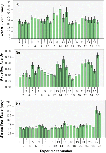
Figure 5. RMS errors for the different interfaces. (a) Visual interface. (b) Navigation compass. (c) Sound guidance. The variation is the standard error of each interface collapsed across all participants and across all related experimental cells shown in .
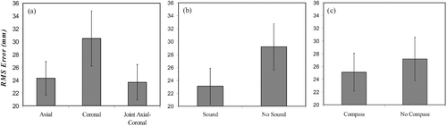
Figure 6. RMS errors for the various interacting factors. (a) Visual-compass interaction. (b) Sound-stereo interaction.
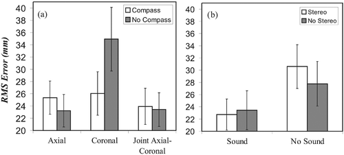
Figure 7. Fraction of measurements recorded inside the tumor for the different interfaces and interaction. (a) Visual interface. (b) Sound guidance. (c) Compass. (d) Visual-sound interaction.
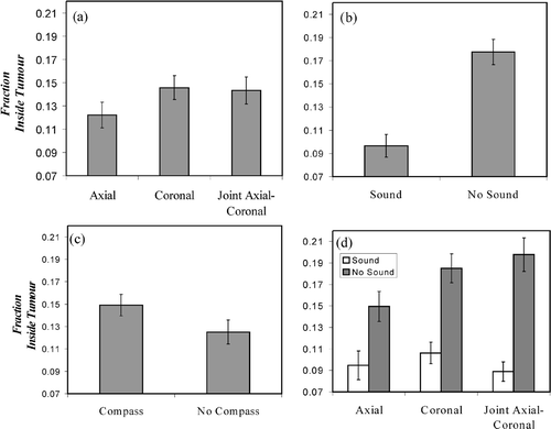
Figure 8. Execution times for the different interfaces. (a) Visual interface. (b) Sound guidance. (c) Stereo vision.
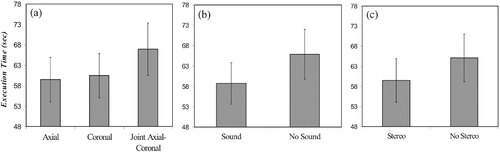
Figure 9. Execution time for the various interacting factors. (a) Visual-stereo interaction. (b) Sound-stereo interaction.
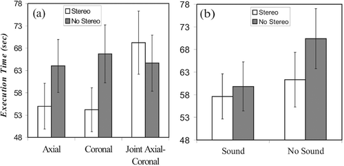
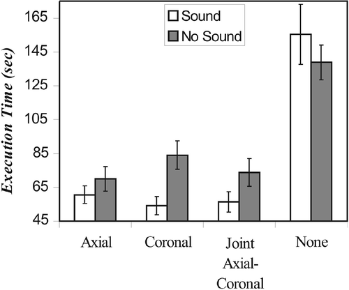
Figure 10. Execution time under “pure” computer-guided conditions for the visual-sound interaction for each of the four visual presentations including no visual presentation (shown as “None”).