Figures & data
Figure 1. Reference points on the lower dental arch. A, B and C are reference points glued to the attached gingiva with an adhesive agent at the right molar, incisal and left molar regions, respectively. [Color version available online.]
![Figure 1. Reference points on the lower dental arch. A, B and C are reference points glued to the attached gingiva with an adhesive agent at the right molar, incisal and left molar regions, respectively. [Color version available online.]](/cms/asset/11af5453-0e81-49fe-b439-5b7367e2e059/icsu_a_225300_f0001_b.gif)
Figure 2. Coordinate system in 3D cephalometric modeling. The reference spheres (A, B and C) are recognized on the lateral and PA cephalometric images and their 3D coordinates are computed. [Color version available online.]
![Figure 2. Coordinate system in 3D cephalometric modeling. The reference spheres (A, B and C) are recognized on the lateral and PA cephalometric images and their 3D coordinates are computed. [Color version available online.]](/cms/asset/28106c24-2115-48a8-96bc-fb267d315c24/icsu_a_225300_f0002_b.gif)
Figure 3. FASTRAK 3D motion tracking and digitizing equipment. (1) System electronics unit (SEU). (2) Transmitter. (3) Box-type receiver. (4) Stylus-type receiver. [Color version available online.]
![Figure 3. FASTRAK 3D motion tracking and digitizing equipment. (1) System electronics unit (SEU). (2) Transmitter. (3) Box-type receiver. (4) Stylus-type receiver. [Color version available online.]](/cms/asset/adce68c8-328c-4fbc-9f18-ad18dcf1b3eb/icsu_a_225300_f0003_b.gif)
Figure 4. Three-dimensional digitizing of centric stops and reference points. A, B and C are the reference spheres impressed on the lower cast model.
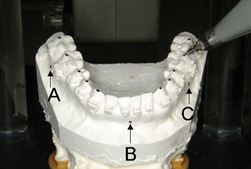
Figure 5. Three-dimensional analysis of centric stops. [Color version available online.]
![Figure 5. Three-dimensional analysis of centric stops. [Color version available online.]](/cms/asset/33be28a4-0e12-46dc-959e-42f9377ba674/icsu_a_225300_f0005_b.gif)
Figure 6. Three-dimensional coordinate system in ManMoS. [Color version available online.]
![Figure 6. Three-dimensional coordinate system in ManMoS. [Color version available online.]](/cms/asset/b35156b3-1054-4e4e-9442-ea90e198d23a/icsu_a_225300_f0006_b.gif)
Figure 7. Three-dimensional wire-frame model available for ManMoS. [Color version available online.]
![Figure 7. Three-dimensional wire-frame model available for ManMoS. [Color version available online.]](/cms/asset/2f392f26-ce06-48ad-ab27-aaa7f8f28a71/icsu_a_225300_f0007_b.gif)
Figure 8. Pilot surgical simulation. [Color version available online.]
![Figure 8. Pilot surgical simulation. [Color version available online.]](/cms/asset/967a146a-e91d-4dd5-a500-21dda8bfc313/icsu_a_225300_f0008_b.gif)
Figure 9. Universal joint simulator. (a) Side view: 1 = universal joint, 2 = transmitter. (b) Superior view of the base section. A box-type receiver is placed on the lower cast model. [Color version available online.]
![Figure 9. Universal joint simulator. (a) Side view: 1 = universal joint, 2 = transmitter. (b) Superior view of the base section. A box-type receiver is placed on the lower cast model. [Color version available online.]](/cms/asset/af7fd3a3-222e-4dd2-b5c3-a25eeec4b1b0/icsu_a_225300_f0009_b.gif)
Figure 10. Digitizing the reference points prior to real-time simulation.
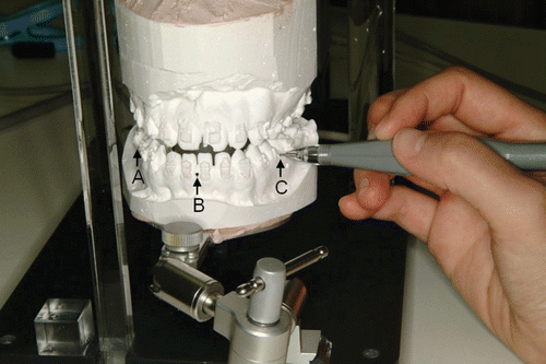
Figure 11. Real-time simulation. [Color version available online.]
![Figure 11. Real-time simulation. [Color version available online.]](/cms/asset/22e82bc8-b86c-4e16-847e-37d6605e3ad5/icsu_a_225300_f0011_b.gif)
Table I. Static noise at ICP.
Table II. Reproducibility.
Figure 12. Fabrication of the occlusal splint. (a) Bite registration. (b) Occlusal wafer splint. [Color version available online.]
![Figure 12. Fabrication of the occlusal splint. (a) Bite registration. (b) Occlusal wafer splint. [Color version available online.]](/cms/asset/b5cf1ad1-e4c8-43ad-b7ed-67bb1b1b85f8/icsu_a_225300_f0012_b.gif)
Figure 13. Soft tissue and skeletal findings in pre-surgical recording. [Color version available online.]
![Figure 13. Soft tissue and skeletal findings in pre-surgical recording. [Color version available online.]](/cms/asset/fe28f73a-64a3-41cb-b887-cfd1ff03be06/icsu_a_225300_f0013_b.gif)
Figure 14. Occlusal findings. (a) Pre-surgical recording. (b) Final mandibular positioning in ManMoS prediction. (c) Fabrication of the occlusal splint (vertical elastic was applied to extrude the upper molar on the right side during the intermaxillary fixation period). (d) Two weeks after surgery (at the end of the intermaxillary fixation). (e) Eight months after surgery (at the end of the post-surgical orthodontics). [Color version available online.]
![Figure 14. Occlusal findings. (a) Pre-surgical recording. (b) Final mandibular positioning in ManMoS prediction. (c) Fabrication of the occlusal splint (vertical elastic was applied to extrude the upper molar on the right side during the intermaxillary fixation period). (d) Two weeks after surgery (at the end of the intermaxillary fixation). (e) Eight months after surgery (at the end of the post-surgical orthodontics). [Color version available online.]](/cms/asset/35cdbc2a-b609-40ff-a0be-1d3e6a02b0c0/icsu_a_225300_f0014_b.gif)
Figure 15. ManMoS simulation with priority given to the occlusal correction. [Color version available online.]
![Figure 15. ManMoS simulation with priority given to the occlusal correction. [Color version available online.]](/cms/asset/7a9cdcd2-25ed-4253-84a5-48674596db71/icsu_a_225300_f0015_b.gif)
Figure 16. ManMoS simulation with priority given to the skeletal correction. [Color version available online.]
![Figure 16. ManMoS simulation with priority given to the skeletal correction. [Color version available online.]](/cms/asset/028f8d02-453b-43c6-8d02-db6864d502a0/icsu_a_225300_f0016_b.gif)
Figure 17. Soft tissue and skeletal findings after 8 months of post-surgical orthodontic treatment. (a) Soft tissue findings. (b) Cephalometric findings. (c) Superimposition of the lateral cephalometric tracings (solid line = 8 months after surgery; dotted line = 1 day after surgery). [Color version available online.]
![Figure 17. Soft tissue and skeletal findings after 8 months of post-surgical orthodontic treatment. (a) Soft tissue findings. (b) Cephalometric findings. (c) Superimposition of the lateral cephalometric tracings (solid line = 8 months after surgery; dotted line = 1 day after surgery). [Color version available online.]](/cms/asset/298f2346-329a-429d-bb7f-c7fea797ecb5/icsu_a_225300_f0017_b.gif)