Figures & data
Figure 1. Simulation of the operating room setting with placement of the cadaveric pelvis in the lateral decubitus position for fluoroscopic image acquisition.
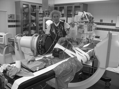
Figure 2. Cadaveric pelvis placed supine for imageless referencing of the pubic tubercle. Note the cup inserter placed for measuring cup position.
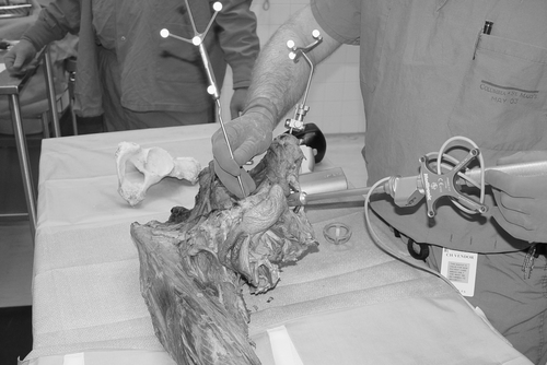
Table I. Measurement precision and variability. (Cp benchmark > 1.3; Cpk benchmark > 2.0)
Figure 3. Position of the more anterior pubic tubercle (A) compared to the pubic symphysis (B).
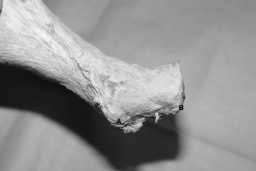
Figure 4. Anatomical reference planes of the pelvis used to calculate anteversion and abduction. [Color version available online.]
![Figure 4. Anatomical reference planes of the pelvis used to calculate anteversion and abduction. [Color version available online.]](/cms/asset/3df45f07-e2ed-49b7-ba0a-ba21c84a1d31/icsu_a_229197_f0004_b.gif)
Table II. Reported precision of reproducibility of different navigation systems using Process Capability Analysis. (Upper and lower specification limits: ±10°)