Figures & data
Table I. Computer-assisted hip resurfacing systems currently on the market.
Figure 1. (a) Dynamic reference base fixation. (b) Field-of-view registration points.
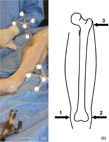
Figure 2. Image acquisition with the zero-dose C-Arm navigation. (a) Preview of frontal view (acquired image in the background). (b) Preview of lateral view prior to second image acquisition. [Color version available online.]
![Figure 2. Image acquisition with the zero-dose C-Arm navigation. (a) Preview of frontal view (acquired image in the background). (b) Preview of lateral view prior to second image acquisition. [Color version available online.]](/cms/asset/69d7367f-cced-4bca-be72-b7a5542d6194/icsu_a_233514_f0002_b.gif)
Figure 3. (a) Safe zone definition. (b) Warning during implant alignment. (c) Final implant position. (d) Offset between planning and result. [Color version available online.]
![Figure 3. (a) Safe zone definition. (b) Warning during implant alignment. (c) Final implant position. (d) Offset between planning and result. [Color version available online.]](/cms/asset/26aa76d0-1601-4b23-8b8d-6f58cc6b9731/icsu_a_233514_f0003_b.gif)
Figure 4. Left: Virtual representation of the tracked pin trajectory superimposed on an acquired X-ray image. Right: Two different points of the tracked pin trajectory in cross-hair representation. [Color version available online.]
![Figure 4. Left: Virtual representation of the tracked pin trajectory superimposed on an acquired X-ray image. Right: Two different points of the tracked pin trajectory in cross-hair representation. [Color version available online.]](/cms/asset/f4de32ac-bba5-47ba-8428-2333ffa1b0ab/icsu_a_233514_f0004_b.gif)
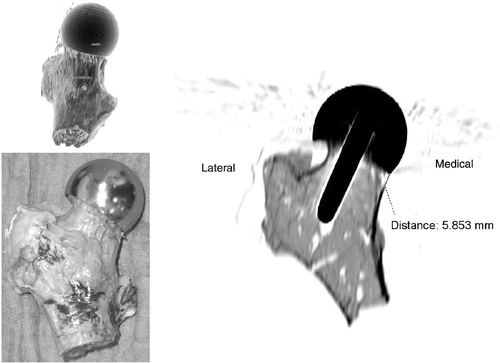
Figure 5. CT evaluation: Implant axis alignment (frontal and lateral view) in relation to femoral neck margins.
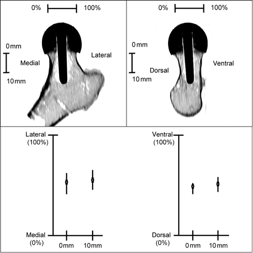
Figure 6. Notched probe: Femoral neck notching on the medial portion.