710
Views
3
CrossRef citations to date
0
Altmetric
Brief Reports

Transepicondylar distal femoral pin placement in computer assisted surgical navigation

Pages 242-246 | Received 20 Aug 2006, Accepted 30 Jan 2007, Published online: 06 Jan 2010

Figures & data

Figure 1. Anatomical specimen demonstrating pins placed medial and lateral in the transepicondylar axis, with a midline pin approximating the “femoral center” landmark which is referenced for the Medtronic StealthStation Treon system. Note the position of the intramedullary rod placement and that all bone cuts, including the femoral notch cut, are not interfered with by this pin placement.
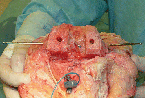
Figure 1. Anatomical specimen demonstrating pins placed medial and lateral in the transepicondylar axis, with a midline pin approximating the “femoral center” landmark which is referenced for the Medtronic StealthStation Treon system. Note the position of the intramedullary rod placement and that all bone cuts, including the femoral notch cut, are not interfered with by this pin placement.

Figure 2. (a) Intraoperative placement of the transepicondylar pins requires dissection of the medial synovial reflection to palpate the medial epicondyle and the supracondylar ridge just cephalad to this point. Pin insertion then parallels the transepicondylar axis and is perpendicular to the AP axis of Whiteside. (b) The femoral array is positioned in the medial side of the lower extremity.
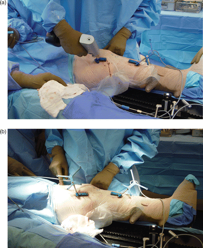
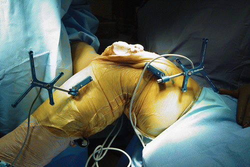
Figure 2. (a) Intraoperative placement of the transepicondylar pins requires dissection of the medial synovial reflection to palpate the medial epicondyle and the supracondylar ridge just cephalad to this point. Pin insertion then parallels the transepicondylar axis and is perpendicular to the AP axis of Whiteside. (b) The femoral array is positioned in the medial side of the lower extremity.

Figure 3. Note the placement of tracking arrays in the medial sagittal plane, which creates an “open field” for the surgical procedure.

Figure 3. Note the placement of tracking arrays in the medial sagittal plane, which creates an “open field” for the surgical procedure.

Reprints and Corporate Permissions

Please note: Selecting permissions does not provide access to the full text of the article, please see our help page How do I view content?

To request a reprint or corporate permissions for this article, please click on the relevant link below:

Academic Permissions

Please note: Selecting permissions does not provide access to the full text of the article, please see our help page How do I view content?

Obtain permissions instantly via Rightslink by clicking on the button below:

If you are unable to obtain permissions via Rightslink, please complete and submit this Permissions form. For more information, please visit our Permissions help page.